Original Contributions
Hum Pathol 1989;20:726-731.
Geographic Variation in the Incidence of Myocardial
Calcification Associated With Acute Myocardial Infarction
SHERMAN BLOOM, MD*
LUDVIK PERIC-GOLIA MD
From the Department of Pathology, The George Washington
University Medical Center, Washington, DC; and the Department of
Pathology, Veterans Administration Medical Center, Salt Lake
City.
*Present address: Department of Pathology, University of
Mississippi Medical Center, Jackson, MS.
Supported by grant no. HL38079 from the National Institutes of
Health and by the Veterans Administration.
Address correspondence and reprint requests to Sherman Bloom, MD,
Department of Pathology, University of Mississippi Medical
Center, 2500 N State St, Jackson, MS 39216.
There is reason to believe that calcium influx into
heart muscle during acute myocardial infarction (AMI) can
aggravate myocyte injury. Furthermore, the degree of such
influx might correlate with the occurrence of microscopic
myocyte calcification observed at autopsy. We have searched for
evidence of myocyte calcification in hearts of patients found
to have AMI at autopsy at the Veterans Administration Medical
Center in Salt Lake City (SLCVA), a region with a low
myocardial infection death rate, and at the George Washington
University Medical Center in Washington, DC (GWUMC), a region
with a high myocardial infection death rate. Of 23 consecutive
cases examined under "blind" conditions at the GWUMC in which
AMI was found, there were 15 instances of cardiac myocyte
calcification observed in von Kossa-stained sections. Not a
single example of myocyte calcification was found in 23
comparable cases at the SLCVA. The basis of this difference in
myocyte calcification is unknown, but may be related to the
fact that the Salt Lake City drinking water contains a higher
level of magnesium, which is known to protect against soft
tissue calcification, than does that of Washington, DC. This
may be the basis for the apparent protection that dietary
magnesium exerts against myocardial infarction
death.
Key words: myocardial infarction,
magnesium, drinking water, geography, myocyte calcification,
metastatic calcification.
Myocardial infarction death rates vary widely as a function of
geography. Some of this variation has been attributed to
variation in dietary intake of magnesium, primarily through
drinking water (1-6). Furthermore, there is an inverse
relationship between the serum magnesium level and the likelihood
of developing a cardiac arrhythmia (7,8), and survival after
acute myocardial infarction (AMI) is increased by the
administration of magnesium (9). These observations are
particularly important in view of the apparent
magnesium-deficient status of a large part of the population in
the United States (10).
In experimental animals, consumption of a magnesium-deficient
diet leads to increased myocardial calcium levels and increased
vulnerability to myocardial necrosis. This necrosis is associated
with myocyte calcification (11,12). In view of the deleterious
effects of calcium on myocyte energy metabolism (13,14), we have
postulated that the increase in myocardial calcium is a critical
determinant of the deleterious effect of magnesium-deficient
diets (15-17). These observations, and the unsubstantiated
impression that AMI-associated myocyte calcification is common in
Washington, DC, but rare in Salt Lake City, led us to hypothesize
that the increased incidence of myocardial infarction death in
areas with low magnesium in the drinking water is associated with
a greater tendency to develop histologically identifiable
myocardial calcification in association with AMI. Such an
increased tendency for calcification would reflect the underlying
abnormality in calcium metabolism hypothesized to be the basis of
the increased AMI death rate in magnesium-poor geographic
regions.
To test the above hypothesis, we compared autopsy material
from patients in two regions where these factors are divergent.
The George Washington University Medical Center in Washington, DC
(GWUMC) was used to represent a high myocardial infarction death
rate area, and the Veterans Administration Medical Center in Salt
Lake City (SLCVA) was used to represent a low myocardial
infarction death rate area. These areas are in the highest and
lowest quintiles of myocardial infarction death rates,
respectively, in the United States (18). We also compared the
mineral composition of the drinking water from these two regions,
using published information and data from water-testing
laboratories.
METHODS
Autopsy reports from both GWUMC and SLCVA for the year 1986
were examined, and all cases in which recent or healed myocardial
necrosis was found were selected for further study. This
consisted of examination of all hematoxylin-eosin-stained
histologic sections taken from the heart. These sections were
obtained from blocks of myocardium, 2 to 4 mm thick, fixed in
phosphate-buffered, 4% formaldehyde at neutral pH. Those patients
in whom microscopy showed evidence of AMI were selected as study
cases. AMI was defined as acute myocyte necrosis evidenced by
loss of cross striations and nuclei, with or without the presence
of contraction bands, and with or without an inflammatory
reaction. Lesions in various stages of repair were also accepted
as showing AMI, including those in which there was evidence of
prior necrosis with inflammation and early repair (granulation
tissue). Healed lesions showing only mature collagen, with or
without chronic inflammatory cells, hemosiderin-laden
macrophages, or dilated capillaries, were not studied further.
Evidence of coronary artery disease was also required: either a
statement that there was at least a 75% reduction in luminal
diameter of a major coronary artery, a statement that coronary
artery narrowing was "severe," or a microscopic slide showing at
least 75% reduction of luminal diameter.
For each case, one slide was selected for staining by the von
Kossa method (19) with hematoxylin-eosin counter staining. This
selection was based on the presence of probable calcium deposits
noted in the hematoxylin-eosin-stained slides. If no such suspect
deposits were found, sections showing the most extensive AMI were
selected, with preference given to sections also showing some
adjacent normal myocardium. Additional slides were prepared from
these blocks, stained by the von Kossa method, and faintly
counter-stained with nuclear fast red. With these sections, von
Kossa-positive deposits were especially conspicuous, but other
morphologic features were poorly stained and more difficult to
identify. Random numbers were assigned to these sections, which
were then examined by one investigator, who scored for the
presence or absence of calcium deposits. The other investigator
examined these same slides, but also had access to
hematoxylin-eosin-stained sections from the same paraffin blocks
as well as von Kossa stained sections counterstained with
hematoxylin-eosin. These other stains were needed to identify the
sites at which the von Kossa-stained material was located.
Although the von Kossa stain for calcium actually stains
phosphate and other precipitated anions, the only known form of
such insoluble deposits in human tissues is the calcium salt.
Therefore, a positive von Kossa stain was considered to be
evidence of calcium depositions We henceforth refer to such von
Kossa-positive material as reflecting calcium deposition.
Histologic preparations, autopsy records, and clinical records
of all cases were examined for evidence of kidney and parathyroid
disease. Cases were identified as having kidney disease if the
serum BUN levels exceeded 40 mg/dL, if the serum creatinine level
exceeded 1.2 mg/dL, or if significant morphologic evidence of
renal parenchymal damage was found. The sex, age, and race of
each patient were also documented. Statistical analyses were
performed on an IBM-AT computer using a commercial software
package (True Epistat, San Antonio, TX). The threshold accepted
for significance was P < .05.
RESULTS
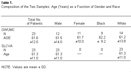
Patients in the GWUMC sample were primarily from the greater
Washington, DC area. There were 14 patients from the District of
Columbia, five patients from adjacent areas in Virginia, and one
patient from an adjacent area in Maryland. In addition, there was
one patient from North Carolina, one patient from Pennsylvania,
and one patient from Pauliabagh, India. For these autopsied
cases, 6.3 ± 0.74 sections taken from the left ventricle
and 4.0 ± 0.69 sections taken from the right ventricle
were examined per case (mean - SD). The GWUMC cases included
males and females and blacks and whites (Table 1).
Patients at the SLCVA were primarily from the central
inter-mountain area. There were seven patients from Salt Lake
City, nine patients from other communities in Utah (six of these
from Salt Lake County), two patients from New Mexico, and one
patient from each of the following states: Idaho, Montana,
Oklahoma, California, and Alaska. All of the SLCVA cases were
white males. For the SLCVA cases, 3.52 ± 0.70 sections
from the left ventricle and 1.41 ± 0.6 sections from the
right ventricle were examined per case.
Following the criteria used in this study, ten cases of kidney
disease were found in the GWUMC sample, while seven cases were
found in the SLCVA sample. In the GWUMC group, there were two
cases of parathyroid hyperplasia and four cases with a malignancy
of some type. These malignancies were metastatic leiomyosarcoma
of the uterus (one case), well-differentiated adenocarcinoma of
the prostate with no demonstrated metastases (one case), renal
cell carcinoma without metastases (one case), and adenocarcinoma
of the prostate with metastases (one case). In the SLCVA cases,
there was one case of parathyroid hyperplasia and no cases of
malignancy. However, there was one case of pheochromocytoma,
thought to be benign and therefore not considered a malignancy.
None of these differences between the SLCVA and GWUMC samples
were significant by Fisher's exact test.
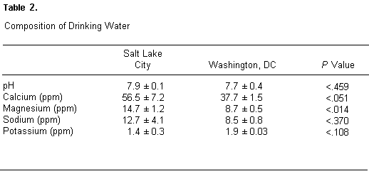
Table 2 shows selected features of the composition of drinking
water in Salt Lake City and the District of Columbia. Mean values
of pH and the content of calcium, magnesium, sodium, and
potassium in the six water sources used in Salt Lake City and the
three water sources used in the District of Columbia are given
for the year 1962. A significant difference in the magnesium
content of these water supplies can be seen (P < .0 14). The
difference in calcium content of the water supplies of the two
areas is not clearly significant (P < .051); calcium, however,
cannot be ruled out as a factor of importance from these data.
Although the values shown in Table 2 are based on samples taken
in 1962, spot checks of data from local water companies in both
Salt Lake City and in the Washington, DC area suggest that these
differences also existed in 1987, but complete contemporary data
for all sources of drinking water in these areas could not be
obtained. The more recent data that is available is given in the
legend to Table 2. These contemporary data support the prior
observation regarding magnesium.
NOTE. Probability values were determined using the
two-tailed t test calculated by a commercial software
package (True Epistat) on an IBM-AT computer. In 1987, the water
content of calcium, magnesium, sodium, and potassium for seven
water distribution sites in the Washington, DC area was 36.5 +/-
9.5, 7.0 +/- 3.5, 16.2 +/- 10.4, and 2.4 +/- 0.4 (mean +/- SD).
In the Salt Lake City area, the corresponding values for 11
distribution sites were 50.0 +/- 25.6, 14.0 +/- 6.8, 12.0 +/-8.9,
and 1.4 +/- 0.6. The differences between these means had
probability values of .322, .037, .450, and .008. These
contemporary data were obtained directly from the local water
districts, and may not be as representative as the data given in
the report by Dufor and Becker (20).
The von Kossa-stained slides revealed a striking difference
between the SLCVA and GWUMC cases. In the former, there was not a
single case of myocardial calcium deposition. In the GWUMC
sample, there were 15 such cases of the 23 cases studied (Table
3). Both observers independently classified these 15 cases as
positive, and all 23 of the SLCVA cases were classified as
negative. There were, however, two cases in the GWUMC sample that
showed von Kossa-positive material that could not be scored as
positive because it was uncertain whether this positive staining
material was within cardiac myocytes. Duplicate sections from the
tissue blocks in question were stained by the von Kossa method
with heinatoxylineosin counterstain and examined by both
observers. Using these sections, both observers agreed that the
von Kossa-positive material was not in cardiac myocytes. In one
case, the von Kossa-positive material was in a mural thrombus,
while in the other case, it was found only in vascular smooth
muscle and endothelial cells.
Table 3.
Frequency of Myocardial Calcification Among Cases at the SLCVA
and the GWUMC
----------------------------------------------------------------

                  With Calcification   Without Calcification
----------------------------------------------------------------

SLCVA                     0                      23
GWUMC                    15                       8
----------------------------------------------------------------

NOTE. The difference between the two samples was highly
significant (P < .00002; two-tailed, Fisher's exact test.)
The distribution of the von Kossa-positive material, when
present, is of some interest. In some cases, von Kossa-positive
granules were found in cardiac myocytes in which cross striations
were clearly visible (Fig 1). In these cases, the stained
material was finely granular and often distributed in such a way
as to accentuate the cross striations. We take it as axiomatic
that cells showing calcium deposits are not viable, but we cannot
explain the preservation of the normal pattern of cross
striations. Further study of these cells was beyond the scope of
the present investigation. In other cases, blotches and granules
of stained material were found within necrotic myocytes (Fig 2).
In yet other cases, positive-staining material was also found in
capillary endothelial cells and in vascular smooth muscle cells
of otherwise normal-appearing small blood vessels (Fig 3). These
differences were not tabulated since it was not always possible
to determine if the calcium-containing cells showed cross
striations, even when the hematoxylin-eosin-counterstained slides
were examined. In one GWUMC case, von Kossa-positive material was
found only in endothelial and vascular smooth muscle cells. This
case was not counted among the 15 positive cases since our
original criteria specified deposits in working cardiac
myocytes.
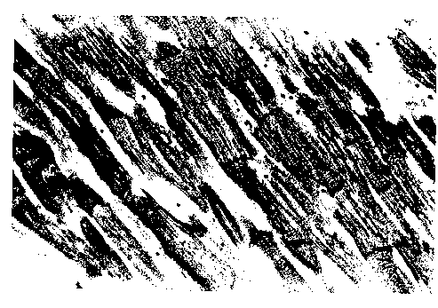
FIGURE 1. A GWUMC patent. Section of myocardium from the
heart of a 64 year-old white man, a resident of the District of
Columbia, who had a healed myocardial intarction and sustained
an AMI less than 1 week before death. Stained by the von Kossa
method and counterstained with hematoxylin-eosin. Although an
AMI was demonstrated elsewhere in the heart of this patient,
this field shows no evidence of myocardial necrosis. Some
myocytes contain granules of von Kossa-positive material that
accentuate the cross-striations.
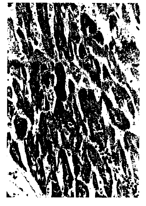
Figure 2. A GWUMC patient. Section of myocardium from the
heart of a 64 year-old black woman who was a resident of the
District of Columbia. Stained by the von Kossa method and
counterstained with hematoxylin-eosin. This field shows the
necrotic myocardium of an AMI judged to be less than 30 hours
old on the basis of history and the morphologic pattern. The
myocytes show loss of nuclei and loss of cross-striations, but
no significant degree of inflammation is yet apparent. Some
cells contain numerous granules, some contain only a few
granules, and some contain no granules of von Kossa-positive
material.
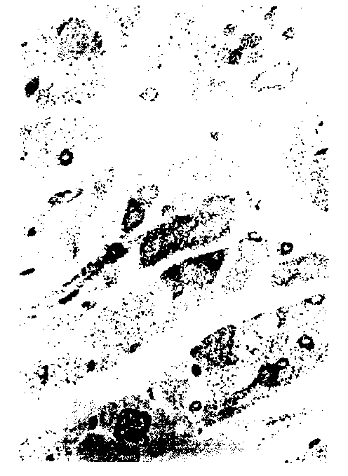
Figure 3. A GWUMC patient. Section of myocardium from the
heart of a 60-year-old white man whose home was in Chapel Hill,
NC, but who was among the GWUMC patient sample. Stained by the
von Kossa method and counterstained with nuclear fast red. This
field shows the cardiac myocytes in cross-section. Some
myocytes contain numerous granules of von Kossa-positive
material. A number of small blood vessels and capillaries also
contain positive-staining material.
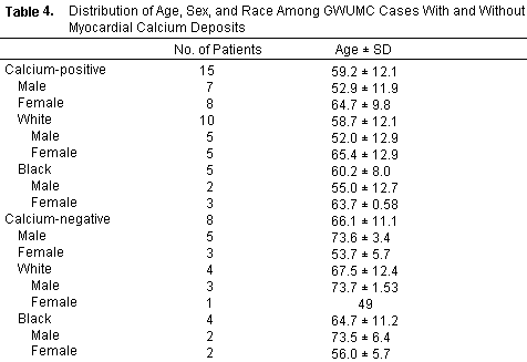
Table 4 shows some characteristics of the GWUMC patient
sample. Comparison of cases with myocyte calcification to those
without such calcification revealed no significant difference
based on race or sex. Analysis of variance (ANOVA) showed no
difference in ages among those with and without calcium deposits,
even when these groups were subdivided by race. When subdivided
by gender, however, ANOVA indicated a clear difference among
groups. This was due to the difference between males who were
positive for myocardial calcification (52.9 ± 11.9 years)
and males who were negative (73.6 ± 3.4 years; P
<.0036, two-tailed). There was no demonstrable difference in
age between positive and negative cases among females (P =
.1045), whites (P = .1354), or blacks (P = .4736). Of the GWUMC
cases that were negative for myocardial calcification, four
listed the District of Columbia as their site of residence and
four listed nearby communities in northern Virginia.
It is possible that the difference in frequency of myocardial
calcification between the two populations studied is due to the
fact that the GWUMC cases were varied in regard to race and sex,
while the SLCVA cases were all white males. There were eight
white males in the GWUMC group, and these had an average age of
60.1 ± 14.9 years. This was not significantly different
from the average age of the 23 SLCVA cases, which were all white
males (61.7 ± 11.1 years). When we only consider the white
male GWUMC cases, we still find a significantly increased
frequency of myocardial calcification compared with the SLCVA
cases (Table 5).
Table 5.
Frequency of Myocardial Calcification Among the White Male SLCVA
and GWUMC Cases
----------------------------------------------------------------

                  With Calcification   Without Calcification
----------------------------------------------------------------

SLCVA                     0                      23
GWUMC                     5                       3
----------------------------------------------------------------

NOTE. The difference between the two samples was highly
significant (P < .00004; two-tailed, Fisher's exact test.)
Table 6 shows the frequency of various ages of infarction as
determined histologically. Such infarct dating is obviously
imprecise, but the data shown indicate that there was no
recognizable difference in infarct age among cases.
Table 6.
Age of Myocardial Infarcts Among SLCVA and GWUMC Cases
----------------------------------------------------------------

                                           GWUMC
Infarct Age      SLCVA      Without Calcium      With Calcium
----------------------------------------------------------------

Hours              0                1                 1
Days              20                8                11
Weeks             11                3                 5
Months            16                7                 8
----------------------------------------------------------------
NOTE. Myocardial infarct age was determined as
"hours" old if there was a significant lesion comprised of
necrotic cardiac myocytes with no associated inflammatory
reaction; as "days" old if there was significant inflammation; as
"weeks" old if there was granulation tissue; and as "months" old
if there was dense scar tissue. Chi square tests showed no
difference between groups shown in this table (P = .692). In
addition, there was no difference between the SLCVA cases and the
combined GWUMC cases (P = .875). The sum of the number of cases
with each age of infarction exceeds the actual number of cases
because in many cases there were infarcts with more than one age.
Among the SLCVA cases, there were only five with infarcts of only
one age; all of these were "days" old. Among the GWUMC cases,
there were seven with infarcts of only one age. Of these, five
were "days" old, one was "weeks" old, and one was "months"
old.
DISCUSSION
It has been known for many years that dystrophic calcification
may affect cardiac myocytes. That is, necrotic cardiac myocytes
may become calcified, especially in patients who have a high
serum calcium level or who are in uremia.(21,22). This, however,
is the first report of geographic variation in the incidence of
AMI-associated myocyte calcification. The GWUMC and SLCVA samples
showed a clear difference in the incidence of AMI-associated
myocardial calcification, but a number of potential confounding
factors are obvious, including a difference in gender and racial
composition. Nevertheless, when only white males were compared,
there was still a highly significant difference in the incidence
of AMI-associated calcification in the two samples, suggesting
that the observed difference was indeed due to a geographic
factor. Even if the difference between the two populations
studied is due to some unrecognized factor, it may be of great
importance. There was no difference in the mean ages of the GWUMC
and SLCVA groups. Within the GWUMC group, there was an unexpected
and striking age difference between those male patients with
AMI-associated myocardial calcification and those without. We
have no explanation for this difference, which was not apparent
among the females.
The fact that three patients in the GWUMC sample showing
myocardial calcification were not from the immediate District of
Columbia area suggests that the geography-associated factor that
determines this effect does not require long-term residency. We
have no information, however, on the water composition in the
communities in which these patients resided, nor do we know how
long they were in the District of Columbia area before they
incurred their AMIs.
Calcification of vascular structures, as noted in several
cases (Fig 3), could also be of considerable significance. It has
been shown that vascular smooth muscle cells show increased tone
when bathed in a medium low in magnesium (23). This may be
related to the inverse correlation between serum magnesium and
systemic blood pressure (24). Von Kossa-positive granules have
been found in the vascular smooth muscle of intramyocardial
arteries of hamsters fed a low-magnesium diet, and this was
associated with fibrinoid necrosis of some arteries (25). These
observations suggest that magnesium deficiency may predispose to
coronary artery spasm, and preliminary studies using experimental
animals support this hypothesis (26).
The present findings raise the possibility that the factors
that influence AMI-associated myocardial calcification may also
play a role in the AMI death rate. Previous investigators have
shown that reduced dietary intake of magnesium is associated with
an increased risk of death caused by myocardial infarction, as
previously discussed, and drinking water in the Salt Lake City
area has a much higher magnesium content than the water in the
Washington, DC area. Furthermore, we have previously shown that
reduced levels of dietary magnesium in experimental animals leads
to reduced levels of serum magnesium and increased myocardial
calcium content, and is associated with increased vulnerability
to ischemic and isoproterenol-induced necrosis (12). This
property of dietary magnesium has led some investigators to refer
to serum magnesium as a physiologic calcium channel blocker (27).
This same mechanism has been proposed as the basis for the
protective effect of magnesium in the preservation of perfused
organs (23).
For the reasons given above, it is tempting to postulate that
we found no myocardial infarction associated calcification in the
Salt Lake City cases because the high magnesium content of their
drinking water led to a low myocardial level of calcium and to a
reduced tendency to deposit calcium at the time of infarction.
The opposite would be true for the Washington, DC area
population. According to this analysis, the calcium-loaded
myocardium of the Washington, DC area patients was more
vulnerable to necrosis because of the metabolic burden associated
with high intracellular levels of calcium (13,29). In both
groups, the sympathetic discharge associated with infarction (30)
could have been expected to increase calcium influx into heart
muscle, but this would have occurred to a greater extent in the
Washington, DC area patients because they lacked the protective
effects conferred by a high dietary level of magnesium. It is
this catecholamine-associated burst of calcium influx,
superimposed on an already increased myocardial level, that is
presumably responsible for the histologically demonstrable
myocardial infarction-associated myocardial calcification in the
Washington, DC area patients, since no such calcification is
found in patients without myocardial infarction.
The results presented here lead us to predict that persons
living in the Salt Lake City area have higher serum magnesium
levels and lower myocardial calcium levels than persons living in
the District of Columbia area. Although the differences may be
small, they may confer a protective effect in myocardial
ischemia.
REFERENCES
1. Neri LC, Johansen HE: Water hardness and cardiovascular
mortality. Ann NY Acad Sci 304:203-219, 1978
2. Karppanen H, Pennanen R, Passinen L: Minerals-coronary
heart disease and sudden death. Adv Cardiol 25:9-24, 1978
3. Johnson CJ, Peterson DR, Smith EK: Myocardial tissue
concentration of Mg and K in men dying suddenly from ischemic
heart disease. Am J Clin Nutr 32:967-970, 1979
4. Punsar S, Karvonen MJ: Drinking water quality and sudden
death: Observations from west and east Finland. Cardiology
64:24-34, 1979
5. Marier JR, Neri LC: Quantifying the role of Mg in the
interrelationship between human mortality/morbidity and water
hardness. Magnesium 4:53-59, 1985
6. Leary WP: Content of magnesium in drinking water and deaths
from ischaemic heart disease in white South Africans. Magnesium
5:150-153, 1986
7. Dyckner T: Serum magnesium in acute myocardial infarction:
Relation to arrhythmias. Acta Med Scand 207:59-66, 1980
8. DeCarli C, Sprouse G, LaRosa JC: Serum Mg levels in
symptomatic atrial fibrillation and their relation to rhythm
control by intravenous digoxin. Am Cardiol 57:956 959, 1986
9. Rasmussen HS, Norregard P, Lindeneg O, et al: Intravenous
magnesium in acute myocardial infarction. Lancet 1:317-322,
1986.
10. Morgan KJ, Stampley GL, Zabik ME, et al: Magnesium and
calcium dietary intakes of the US population. J Am Coll Nutr
4:195-206, 1985
11. Heggtveit HA, Herman L, Mishra RK: Cardiac necrosis and
calcification in experimental Mg deficiency. Ann NY Acad Sci
162:758-774, 1964
12. Bloom S: Magnesium deficiency cardiomyopathy. Am J
Cardiovasc Pathol 2:7-17, 1988
13. Bloom S: Reversible and irreversible injury: Calcium as a
major determinant, in Balazs T (ed): Cardiac Toxicology. Boca
Raton, FL, CRC Press, 1981, pp 179-199
14. Kitakaze M, Weisman HF, Marban E: Contractile dysfunction
and ATP depletion after transient calcium overload in perfused
ferret hearts. Circulation 79:685-695, 1988
15. Chang C, Varghese J, Downey J, et al: Magnesium deficiency
and myocardial infarct size in the dog. J Am Coll Cardiol
5:280-289, 1985
16. Chang C, Bloom S: Interrelationship of dietary Mg intake
and electrolyte homeostasis in hamsters. j Am Coll Nutr
4:173-185, 1985
17. Bloom S: Effects of Mg deficiency on the pathogenesis of
myocardial infarction. Magnesium 5:154-164, 1986
18. Moriyama I, Krueger D, Stamler J: Cardiovascular Diseases
in the United States. Cambridge, MA, Harvard University, 1971, pp
49-118
19. Pearse AGE: Histochemistry: Theoretical and Applied.
Baltimore, Williams & Wilkins, 1972, pp 1138-1139
20. Durfor CN, Becker E: Public Water Supplies of the 100
Largest Cities in the United States, 1962. Geological Survey
Water-Supply Paper. Washington, DC, US Government Printing
Office, 1965, pp 136 and 346
21. Gore I, Arons W: Calcification of the myocardium: A
pathologic study of thirteen cases. Arch Pathol 48:1-12, 1949
22. Scotti TM: Heart, in Anderson WAD, Kissane JM (eds):
Pathology, vol 1. St Louis, Mosby, 1977, pp 745-746
23. Turlapaty PDM, Altura BM: Magnesium deficiency produces
spasms of coronary arteries: Relationship to etiology of sudden
death of ischemic heart disease. Science 208:198-200, 1980
24. Dyckner T, Wester P: Effect of magnesium on blood
pressure. Br Med J 286:1847-1849, 1983
25. Bloom S: Coronary arterial lesions in Mg-deficient
hamsters. Magnesium 4:82-95, 1985
26. Yeager JC, Masters TN: Thermographic evidence that an
ergonovine-induced coronary artery spasm can be produced in the
dog by acutely lowering plasma Mg. Magnesium 1:95-103, 1982
27. Levine BS, Coburn JW: Mg, the mimic/antagonist of calcium.
N Engl J Med 310:1253-1255, 1984
28. Shattock MJ, Hearse DJ, Fry CH: The ionic basis of the
anti-ischemic and anti-arrhythmic properties of magnesium in the
heart. J Am Coll Nutr 6:27-33, 1987
29. Fleckenstein A: Specific inhibitors and promoters of
calcium action in the excitation-contraction coupling of heart
muscle and their role in the prevention or production of
myocardial lesion, in Harris P, Opie L (eds): Calcium and the
Heart. San Diego, Academic, 1971, pp 135-188
30. Willerson JT, Buja LM: Beta-adrenergic mechanisms during
severe myocardial ischemia and evolving infarction. Postgrad Med
1988, pp 27-32
This page was first uploaded to The Magnesium Web Site on June
12, 1996
http://www.mgwater.com/


