MAGNESIUM DEFICIENCY IN THE PATHOGENESIS OF DISEASE
Early Roots of Cardiovascular, Skeletal
and Renal Abnormalities
Goldwater Memorial Hospital
New York University Medical Center
New York, New York
1980
(include the word "jacket" to search only in this book)
| Jacket | Preface | Contents | Introduction (Chapter 1) |
Chapter: | 2 | 3 | 4 | 5 | 6 | 7 | 8 | 9 | 10 | 11 | 12 | 13 | 14 |
| Appendix | Bibliography (A-D), (E-K),
(L-R), (S-Z) |
Part II: Chapter 9
MAGNESIUM DEFICIENCY IN THE PATHOGENESIS OF CARDIOVASCULAR DISEASES
9
Magnesium Deficiency and Cardiac Dysrhythmia
9.1. Electrocardiographic Changes of Experimental Magnesium Deficiency
In the early subacute magnesium-deficiency study of Kruse et al. (1932), convulsions were produced in 86% of the rats by the 18th day, with death occurring after one or more convulsions in 93%. Tachycardia was manifest during the preconvulsive period, and bradycardia with marked arrhythmia just before the convulsions started. Greenberg and Tufts (1938) confirmed these findings, and showed additionally that ECGs, taken while the rats were unconscious from the convulsive seizures, revealed a sinoauricular block, with occasional skipped and ventricular beats. Of 10 rats with less severe magnesium deficiency, such that despite manifest nervousness only one developed convulsions, seven survived long enough to have ECGs recorded the day before sacrifice on day 62. These rats exhibited little change in heart rates (which were slightly slower than were those of control rats on the same diet to which magnesium had been added) but had lengthened P-R intervals. Five of the seven surviving deficient rats had additional ECG abnormalities: Three had numerous extrasystoles, two had abnormally high takeoff of the ST segment in lead III, one with partial heart block and one with auricular extrasystoles.
Production of magnesium deficiency (average serum magnesium = 0.4 mEq/liter) in young dogs, with a diet similar to that used by Kruse et al. (1932), produced no significant difference from control heart rate (Syllm-Rapoport et al., 1962). There was a highly significant shortening of the atrioventricular conduction time (P-Q interval) and of the intraventricular conduction time (QRS in Lead II). There was some prolongation of the electrical systole (QT interval). There was an increased incidence of negative T waves in leads I, II, and III that was statistically significant in lead III. The voltage of the negative T waves in leads I, II, and III was statistically significant in lead III. The voltage of the negative T waves in leads I and II was almost half that of controls. Striking inversion of the T waves was seen in several of the deficient animals. Comparably severe magnesium deficiency, produced with a semisynthetic diet, and that produced severe hypomagnesemia (< 0.5 mEq/liter), but no significant effect on serum potassium or calcium), and that caused arterial and multifocal myocardial lesions, was also associated with ECG abnormalities (Wener et al., 1964). These dogs developed sinus tachycardia, but little difference from control PR, QT, or QRS intervals. There was frequent occurrence, as in the previous group of dogs, of T-wave abnormalities: flattened or inverted T waves, especially in leads III, aVL, and V. They also had consistent RST-segment depression.
Subacute magnesium deficiency for three months in puppies that resulted in irritability and occasional convulsions also resulted in marked sinus tachycardia, peaking of the T waves, and ST-segment depression (Vitale et al., 1961). These dogs also became more susceptible to digitalis toxicity. These investigators pointed out the relationship of these ECG changes to the magnesium-induced shift in potassium. Although the magnesium-deficient dogs also developed hypokalemia, Vitale et al. (1961) referred to the loss of intracellular potassium that results from magnesium-deficient interference with mitochondrial enzymatic activity. They speculated that there might be a relatively greater decrease in intracellular than plasma potassium with a relative hyperkalemia. In support of this premise, was the peaked T wave of their magnesium-deficient dogs that resembled that seen in hyperkalemia. Ono (1962) confirmed these findings with young dogs maintained for four months on the same diet as used by Vitale and his co-workers (1961). He, too, found peaking of the T waves, especially in lead VR, when the serum magnesium levels decreased to 0.7 mEq/liter, with concomitant falls in serum potassium. Depression of the ST segment in limb or chest lead appeared with serum magnesium levels below 0.8 mEq/liter. The P-R interval increased slightly as the hypokalemia worsened. The Q-T interval remained almost normal. There were also occasional premature contractions. Comparable changes were produced by magnesium deficiency in monkeys (Vitale et al., 1963), except for bradycardia and elevated ST segment in severely deficient monkeys. The peaking of the T waves and the ST segment changes were comparable to those seen in hyperkalemia, even though the animals had hypokalemia. This group then tested their original postulate that there might be a local relative hyperkalemia (of the extracellular/intracellular potassium concentration) in the magnesium-deficient heart (Seta et al., 1965, 1966). Rats fed diets low in both magnesium and potassium had substantial reductions in myocardial potassium and magnesium levels. Rats on a low magnesium, adequate potassium intake had almost normal serum potassium level, but markedly subnormal myocardial potassium (Seta et al., 1965), supporting the premise that the hyperkalemia-like ECG of relatively early magnesium deficiency reflects local relative hyperkalemia. Electrocardiographic changes were observed at two-week intervals: T-wave peaking developed within two weeks of instituting the magnesium-deficient diet (best seen in the left precordial unipolar lead). QRS widening and tachycardia were additional early changes (Seta et al., 1966). ST segment depression, ventricular premature beats, and bigeminal rhythm were also seen in some of the dogs. The ECG changes of dogs deficient in both magnesium and potassium resembled those of potassium deficiency, but the terminal T-wave inversion was more marked. The P-R interval and the QRS segment were prolonged, and there was slight ST-segment depression. The heart rate, unlike that of the animals that were deficient in magnesium but not in potassium, did not change. Dogs that were kept on the magnesium-deficient diet for nine months developed an ECG pattern indistinguishable from that of doubly deficient dogs after two months (Seta et al., 1966)
Electrocardiographic changes, like those of subacute experimental magnesium deficiency have been reported from studies of cattle pastured on land low in available magnesium (Willers et al., 1965). The ECG criteria for detection of the disease termed "bovine arteriosclerosis" are tented T waves, prolonged QRS interval, and elevated ST segments. The investigators noted that such events in man are associated with hyperkalemia, endocardial thickening, and conduction system-interference. Autopsy reports (of cattle from the herd tested electrocardiographically) showed that endocardial thickening and coronary calcific arteriosclerosis were characteristic (Lynd et al., 1965). Larvor et al. (1964a) found that magnesium-deficient calves had tachycardia and shortened PQ intervals. One calf that developed myocardial degenerative changes had had a diphastic T wave. Reference to the conduction disturbance recalls the early microscopic study of magnesium-deficient calves that showed not only endocardial plaques and fibroelastosis and myocardial necrosis, but lesions of the Purkinje fibers (Moore et al., 1936; 1938; Arnold and Fincham, 1950). It also recalls the evidence that the interventricular septum has the greatest avidity for magnesium (Glaser and Brant, 1959; Glaser and Gibbs, 1962; Lazzara et al., 1963; Burch et al., 1965). Thus, the dysrhythmias of magnesium deficiency probably reflect high magnesium requirements of the conduction system, and secondary potassium shifts out of the myocardial cells.
Acute sudden magnesium depletion by hemodialysis has not produced as significant ECG alterations as have subacute or chronic deficiencies. Danzig and Walker (1955) depleted dogs of magnesium over a six-hour period by dialysis, using a magnesium-free, but otherwise physiologically constituted dialysate. The ECGs s the end of dialysis, when the plasma magnesium was 0.34-0.70 mEq/liter showed an increased heart rate and decreased QT interval. Comparable reduction in plasma magnesium during dialysis for 2 1/2 hours caused only 15% increase in rate and slight decrease in PR and QT intervals (Grantham et al., 1960)
Baby pigs, on a synthetic milk diet that was severely deficient in magnesium, developed bradycardia, increased R and T wave potentials, and inverted T waves in the standard leads (Miller et al., 1964a). Moderate magnesium deficiency resulted in tachycardia with a normal R-wave potential. Acute calcium deficiency also produced bradycardia, with a lengthened ST interval.
Bajpai et al. (1978) have correlated the ECG changes produced by hypomagnesemia in rats with abnormalities in mitochondrial oxidative phosphorylation. They confirmed the significant reduction of the P-, QRS-, and T-wave voltages of magnesium deficiency, and attributed the changes to decreased energy production associated with the decreased oxidative phosphorylation. They propose that magnesium deficiency reduces the amount of current transmitted from cell to cell, as a result of increased resistance in the intercellular connections (desmosomes) as these membrane structures swell [similarly to the swelling of the plasma membranes of magnesium deficient erythrocytes (Elin, 1978)].
It might be that the severe forms of magnesium deficiency that are associated with bradycardia and depressed, prolonged ST segments might reflect concomitant hypocalcemia and hypokalemia (Fig 9-1B; Seelig 1969a). The ECGs of the less severe, subacute, or chronic magnesium deficiencies resemble not only that of hyperkalemia but also that of hypercalcemia, or a combination thereof (Fig. 9-lA). Thus, the magnesium-deficiency ECGs reflect also the concomitant or resultant abnormalities of the other two cations that affect the cardiac conduction system, and not the magnesium status alone.
It is provocative that the ECG of magnesium deficiency also resembles that seen in myocardial ischemia of coronary insufficiency: flattened, inverted, or peaked T waves and ST depression, as well as abnormally long QT interval. Its ST depression and T-wave inversion also resemble the ECG of subendocardial infarction, an interesting point, since the small intramural coronaries have been shown to be most compromised in magnesium-deficient animals, and the subendocardial area is most susceptible to ischemia under such circumstances. The presence of endocardial fibroelastosis in infants with perinatal factors that increase the risk of hypomagnesemia is further suggestive evidence that magnesium deficiency might be contributory to the infantile cardiovascular diseases discussed earlier, and that its loss from the heart might be a factor in the ischemic ECG.
9.2. Magnesium Interrelations with Other Factors in Cardiac Rhythmicity
Basic to the role of magnesium in maintaining or restoring normal cardiac rhythmicity and in preventing hyperexcitability is its role in maintaining intracellular accumulation of potassium against a concentration gradient, and in counteracting excess calcium influx.
9.2.1 Magnesium/Potassium in Cardiac Rhythmicity
DeCarvalho (1965) has reviewed the factors controlling electrical activity in heart muscle, which is strongly dependent on the electrolyte balance between the cell and its environment. Potassium ions are particularly important in the genesis of cardiac transmembrane potentials, and affect the cardiac rate, impulse conduction, excitability threshold and refractoriness. He considered the influence of alterations in extracellular potassium concentrations on the polarity of the myocardial cell and on transmission of impulses in the special conducting system of the heart. High concentrations of extracellular potassium depress impulse propagation in the atrium and in the His-Purkinje system; low extracellular concentrations depress the atrioventricular (A-V) nodal area, A-V block ensuing with very low (1.5 mM) extracellular potassium levels. Under normal circumstances, potassium is extruded from the myocardial cell during systole. Its return during diastole is an energy-dependent process, since it entails transport against a concentration gradient (Raab, 1969).
The active transport of potassium into, and sodium out of the myocardial cell is dependent on the integrity of the mitochondrial enzyme system. Baltscheffsky 1956, 1957) first postulated that magnesium plays a specific role in the respiratory control of the mitochondrion, since it is a cofactor in the oxidative phosphorylation reactions. Without magnesium, the respiratory rate decreases: there is "uncoupling" of oxidative phosphorylation. He also suggested that magnesium is essential to mitochondrial integrity. A. Schwartz (1971/1972) diagrammed the structure and functions of the mitochondrion which contain magnesium-dependent enzyme systems of the Krebs cycle (and provide most of the energy requirements, as well as those controlling oxidative phosphorylation). Lehninger (1962) showed that the mitochondrial functions are responsible for electrolyte and water transport. Aikawa 1965) reviewed the data on the enzymatic importance of mitochondria and hypothesized that magnesium is essential for the metabolic activity of all subcellular particles. He speculated that there might be an "unknown carrier molecule" that might be involved in the active transport of the magnesium ion across the inner membrane of the mitochondrion, such as has been identified in cardiac mitochondria (Blondin, 1975; Green et al., 1975).
The importance of oxidative phosphorylation, particularly in Mg-activated membrane ATPase (that is vital in electrolyte transport), was first shown in noncardiac tissue such as nerves, brain, kidney, and erythrocyte membranes (Skou, 1957, 1960, 1962; Post et al., 1960; Dunham and Glynn, 1961; Whang and Welt, 1963; Welt and Tostesen, 1964; Welt, 1964), and in cardiac and other mitochondria and microsomes (Nakamura et al., 1961; DiGiorgio et al., 1962; Auditore, 1962; Auditore and Murray, 1963; Vitale et al., 1963; Schwartz, 1962; Schwartz and Laseter, 1964).
The loss of myocardial potassium in magnesium-deficient animals was first attributed by Vitale and his colleagues (1975a,b) to the uncoupling of oxidative phosphorylation. The myocardial cells (which are vulnerable to magnesium loss because of the high percentage exchangeability of their magnesium, not only lose the capacity to accumulate potassium against a concentration gradient and pump out sodium, but show concomitant mitochondrial disorganization, a not surprising correlation in view of the dependence on the integrity of the mitochondrial enzymes systems for active electrolyte transport.
9.2.2. Catecholamine/Magnesium/Potassium Interrelationships
Epinephrine, whether injected (W. Robertson and Peyser, 1951) or secreted as a result of stress (Raab et al., 1968), causes a decrease in the intracellular potassium/sodium ratio. Using the potent β-adrenergic agonist (isoproterenol), Lehr et al. (1966) showed that the earliest myocardial electrolyte changes were significant decreases in magnesium and phosphorus and an increase in calcium. These changes were noted at 3 hours after injection of the catecholamine, in association with mild microscopic evidence of necrosis, but no significant changes in potassium or sodium. There was a significant rise in myocardial sodium and a minor fall in myocardial potassium at 12 hours, at which time the abnormalities in magnesium, calcium, and phosphorus were greater and all of the rats had severe myocardial necrosis.
These observations may help to explain the marked similarity of the ECG produced by excessive sympathomimetic substances (Raab, l943b) and those produced by nutritional magnesium depletion, whether produced in the experimental animal or as a result of chronic alcoholism or protein calorie malnutrition (Seelig, l969a). In both there can be elevation or depression of the ST segment, abnormalities of the T wave, ranging from a high pointed shape to inversion and prolongation of the QRS or QT interval. Raab (1943b) pointed out that the epinephrine-ECG pattern reflects the relative hypoxia produced as oxygen consumption exceeds oxygen supply. It is of interest that chronic magnesium deficiency has been shown to cause luminal narrowing of intramyocardial coronary arteries and also to interfere with normal mitochondrial respiration supra vide).
The DOCA saline pretreated rats that showed myocardial magnesium depletion and that died of ventricular fibrillation 15 to 30 minutes after minimal doses (60-100 µg/kg) of isoproterenol, developed auricular and ventricular arrhythmias, progressing to fibrillation as the β-agonist dosage was raised. Epinephrine, as α- and β-agonist, elicited arrhythmias and ventricular fibrillation less consistently, and only when α-adrenergic receptors were blocked (Guideri et al., 1974).
It is surprising that the ECG changes and myocardial damage produced in rabbits stressed by being kept in a vertical position were significantly protected against by oral administration of magnesium chloride (1 g/kg) twice daily, whereas those injected with epinephrine (0.2 mg/kg) intravenously were not similarly protected by MgCl2 (Pokk, 1971/1973). The catecholamine-injected rabbits had been given an unspecified amount of the magnesium every five days before the injection and thereafter. Perhaps the amount given was insufficient to achieve the amelioration of ECG and myocardial changes that was seen in the stressed, magnesium-dosed rabbits. The ECG changes included bradycardia, large R waves, and depressed ST segments.
9.2.3. Postinfarction/Catecholamine/Free Fatty Acid/Magnesium Interrelationships with Arrhythmia
It has been shown that blood and urine catecholamine levels are increased in patients who are severely ill after a heart attack and the catecholamines have been implicated in the postinfarction arrhythmias (Gazes et al., 1959; Richardson et al., 1960; Valori et al., 1967; McDonald et al., 1969; Editorial, Lancet, 1969a). High levels of circulating free fatty acids have also been implicated in postinfarction arrhythmias (Kurien and Oliver, 1966; Kurien et al., 1969, 1971), and the two findings have been correlated by some, in view of the catecholamines' lipolytic effects McDona1d et al., 1969; Editorial, Lancet, 1969b)
When corn oil was added to the diet of sodium phosphate mineralocorticoid treated (ESCN) rat, it developed infarctlike myocardial lesions and electrocardiographic abnormalities that were similar to those produced by magnesium deficiency in association with local relative hyperkalemia (Vitale et al., 1961, 1963; Seta et al., 1966): There was prolongation of the PR and QRS segments, low voltage, and peaking of the T wave, with atrial fibrillation and conduction abnormalities that developed at about the time that necrosis became visible (Varga et al., 1970). Amiloride protected against the severe ECG changes, but tachycardia persisted and the amplitude of the PR and S waves remained elevated. Cardiac necrosis was almost completely prevented. Serum and myocardial electrolyte analyses of rats sacrificed on the fifth day of study suggest that in this model, the amiloride protection might have been mediated by protection against myocardial necrosis closely resembling those of his electrolyte-steroid cardiac-necrosis (ESCN) experimental model in that all produce extensive, usually multifocal myocardial necrosis. Excessive concentrations of epinephrinelike substance in the heart of a young athlete who had died suddenly (Raab, 1943a), and in hearts of patients who had died with angina pectoris and other cardiac dysfunctions (Raab, 1943b), and the similarity of the ECG changes of patients with IHD to those of animals or humans given epinephrine, led Raab to consider stress-induced hormonal (catecholamine and corticosteroid) excess as basic to the disorder he termed cardiac "dysionism." He observed that major shifts in myocardial electrolytes can lead to disturbances in cardiac rhythm, contractility, structure, and ultimately to cell necrosis. His emphasis was on the depletion of intracellular potassium, but he observed that this was usually paralleled by loss of glycogen and magnesium and by entry of sodium into the myocardial cells.
When cardiac function is inadequate for the load capacity of the heart, relative ischemia develops, which can be manifested by angina, sudden death from arrhythmia, or congestive failure. Hochrein and Lossnitzer (1969) have pointed out that when there is hypoxic cardiac dysfunction or failure, the myocardial metabolism is characterized by shift toward anaerobic from aerobic metabolism, with loss of magnesium and potassium and gain of sodium chloride and water (Fig. 9-2A), with resultant myocardial edema and decreased energy production for the amount of oxygen consumed. A similar pattern results from cardiac overload, except that there is first increased lactate consumption with intensified glycolysis, and subsequent inhibition of glycolytic metabolism, again with loss of myocardial magnesium and potassium (Fig. 9-2B).
On the other hand, anoxia itself causes loss of magnesium from the myocardium, as well as increased myocardial lipid accumulation (Review, Opie, 1968). which can further decrease the available magnesium. Even venous occlusion of the arm with a blood pressure cuff causes egress of magnesium from the cells, as reflected by immediate rise in local serum magnesium levels (Whang and Wagner. 1966; Nielsen, 1969). Thus, the similarities between the ECGs of coronary insufficiency and of magnesium deficiency are not surprising. The changes that occur with treatment of the cardiac disease, particularly that increase myocardial calcium and decrease myocardial magnesium (i.e., cardiotonics) and that intensify potassium loss (i.e., diuretics), to which magnesium deficiency contributes (Review: Seelig. 1972) can account for the wide variety of ECG changes seen with cardiac ischemia and decompensation and at different stages of magnesium deficiency.
9.2.4. Blood Primes for Extracorporeal Circulation
The use of acid citrate dextrose (ACD) solution for preserving blood without coagulation is known to remove ionized calcium, and thus calcium is usually added. However, its equal binding capacity for magnesium has not been as widely appreciated, with resultant production of arrhythmias during exchange transfusion in infancy and in open-heart surgery. Killen et al. (1971) have shown that total and ionized magnesium levels dropped to below 1.5 mEq/liter and to about 0.5 mEq/liter, respectively, within two hours of the infusion in dogs. Heparin has been recommended as an anticoagulant, to avoid this problem, but Romero et al. (1973) performed cardiopulmonary bypass using heparinized blood in dogs, and found drop of serum magnesium from a pre-bypass level of 1.6 to 1.2 mEq/liter, which was sustained for the two hours of the bypass and for the hour of observation thereafter. Thus, to avoid the arrhythmias of exchange transfusion and of open- heart surgery, addition of magnesium to the prime is recommended. That the optimal magnesium concentration in the infusate or blood prime might be considerably higher than the physiological concentration, as suggested by the few who have written reports recommending the clinical use of magnesium during open-heart surgery, is indicated by the study of Hearse et al. (1978). Using a rat heart model of cardiopulmonary bypass, they showed magnesium to be the single most effective component of any infusate tested. The concentration at which maximal protective activity was achieved was 15 mmol/liter. Increasing the magnesium concentration from 0 to 15 mmol/liter produced a progressive and significant improvement in the recovery of function during the reperfusion. There was a striking increase in protection between 0 and 2.4 mmol/liter and another at 15 mmol/liter; thereafter the protective effect declined with increasing magnesium concentrations (Table 9-1).
9.3. Magnesium Deficiency in Clinical Arrhythmia
ECG abnormalities (similar to those seen in magnesium-deficient animals or in animals subjected to stress or given hormonal, drug, and electrolyte challenges that produce loss of myocardial magnesium) have been seen in several clinical conditions that have caused magnesium depletion. Many conditions have been listed as associated with hypokalemic or hypocalcemic ECGs, or both (Surawicz and Lepeschkin, 1953; Judge, 1968; Fletcher et al., 1967; Table 9-2). It is of interest that all of these conditions have been associated with magnesium deficiency: hypomagnesemia, low tissue levels, both, or low tissue levels but high serum levels. Electrocardiograms have been recorded while volunteers were depleted of magnesium. Among the diseases in which abnormal ECGs have been recorded, the most acute situations, those that produce sudden and severe hypomagnesemia, often in association with preceding or concurrent stress, are those during which citrated blood is used. Exchange transfusions of infants, open heart surgery, and extensive surgical procedures or repair of blood loss secondary to serious trauma, are the most obvious examples. The first recognition of the risk of hypomagnesemia and associated arrhythmia in patients receiving long-term intravenous infusions was reported by Flink et al. in 1954. The following years this group called attention to the hypomagnesemia of alcohol abuse and diuretic overuse (Flink, 1956; McCollister et al. 1958), both agents that have been associated, as well, with arrhythmias. Infants with "primary" or "idiopathic" myocardial diseases have also been found to have ECG abnormalities resembling those of magnesium deficiency.
9.3.1. Experimental Magnesium Deficiency (Man)
Two normal men were fed diets low in magnesium (1-2.5 mM/day; or 2-5 mEq/day or 25-60 mg/day) and high in calcium for 39 and 48 days, respectively, and were given intravenous infusions of sodium and potassium sulfate to augment renal magnesium loss. One, who developed hypokalemic alkalosis on day 46, developed a hypokalemic ECG, despite a potassium intake of more than 40 mM/day. The plasma potassium rose and the ECG reverted to normal during magnesium repletion (Dunn and Walser, 1966). Seven patients who had had radical head and neck surgery for carcinoma, and thus could be kept on a controlled magnesium-deficient liquid diet for prolonged periods, were more severely depleted of magnesium (Shils, 1969a). Their daily magnesium intakes were 0.5 to 0.8 mEq (60 to 10 mg) for 42 to 266 days. Serial ECGs were obtained on all subjects. Three, who had been depleted for 42, 104, and 117 days, developed changes in the T waves consisting of broadening and decreased amplitude (or occasionally inversion), U waves, and slight prolongation of the QT interval. Two of these patients also had decreased voltage and one had some shortening of the ST segment. A fourth patient had a prolonged QT interval. These changes were associated with low levels of magnesium, calcium and potassium. Only one patient with severe electrolyte changes had no ECG abnormality. The two patients with the least disturbance in electrolytes had no significant ECG changes. It is noteworthy that during the early period of magnesium repletion, two of the patients' low serum potassium and calcium persisted even though their serum magnesium levels had become normal. Their ECGs remained abnormal until later in magnesium-repletion period, at a time coinciding with restoration of normal serum calcium and potassium levels. Possibly this reflects correction of the ECG when the body stores (including cardiac levels) of magnesium were sufficiently repleted to permit restoration of normal mitochondrial and parathyroid function, without which inability to maintain normal potassium and calcium levels is not surprising, despite their supplementation.
9.3.2. Electrocardiographic Changes with Use of ACD Blood
9.3.2.1. Exchange Transfusion
The evidence that exchange transfusion with ACD blood has produced acute magnesium deficiency in newborn infants has been discussed earlier. The most consistent change associated with a drop in serum ionized magnesium to below 0.8 mEq/liter, was characterized by a flat T wave (Bajpai et al., 1971, 1972). A similar tracing was seen in another infant, who was unsuccessfully treated with calcium gluconate for hypocalcemic seizures, noted first six days after he had undergone an exchange transfusion (Dooling and Stern, 1967). A flat T wave was noted in the third week of life when, despite continuation of calcium therapy and sodium bicarbonate treatment of his acidosis, the baby continued to suffer disastrous seizures, which were finally attributed to his concomitant hypomagnesemia (0.6 mEq/liter), reported first from blood taken on the 11th day of life. His tremulousness and seizures ceased in response to 0.25 ml of 50% magnesium sulfate every six hours (providing 25 mEq in 24 hours), but his serum magnesium remained low (0.76 mEq/ liter); it was at this time (three days after the magnesium therapy had been started) that the abnormal ECG was first observed. The infant required 0.5 ml of 50% magnesium sulfate intramuscularly every eight hours to bring his serum magnesium up to 1.6 mEq/liter and to correct the flat T waves. In a short communication, Rosefsky (1972) reported that a premature infant, who had required several exchange transfusions, and who had been given calcium prophylactically, developed arrhythmia with many ectopic ventricular beats during his fourth transfusion. This infant responded to slow intravenous injection of magnesium sulfate (25 to 50 mg/kg) within five minutes, with return of rhythm to normal. When the ectopic beats recurred on subsequent exchange transfusions, intravenous magnesium therapy again rapidly restored the rhythm to normal. ECG changes: high P waves, ST depression, and flat or inverted T waves, are common during exchange transfusions (Robinson and Bathe, 1963). Citrate-lowered ionized magnesium (Bajpai et al., 1967a,b) has been implicated in the high sudden-death rate during this procedure (Editorial, Canad Med Assoc J, 1967).
9.3.2.2. Open-Heart Surgery
The use of ACD blood in the pump oxygenator prime during cardiopulmonary bypass (performed during hypothermic or normothermic anoxic arrest with ventricular fibrillation) has been associated with even more arrhythmias. Scheinman et al. (1969) investigated the pre- and postoperative magnesium status of 17 adult patients undergoing intracardiac operative procedures, after they had observed classic neuromuscular signs of acute magnesium depletion in a patient soon after cardiopulmonary bypass. Levels of three of the patients could not be compared because they had remained in persistent fibrillation, despite standard therapy (including multiple attempts at internal defibrillation) until they were given an intravenous bolus of magnesium sulfate. Of the remaining 14, 12 exhibited drops in serum magnesium from 1.65 ± 0.10 mEq/liter before surgery to 1.07 ± 0.01 mEq/liter after surgery. Despite the hypomagnesemia, no gross neuromuscular abnormalities were noted. This group of investigators then compared those patients with another group of eight, who had magnesium (2 mEq/liter) added to the pump prime (R. Sullivan et al., 1969; Scheinman et al., 1971/1973). Although there was no apparent relationship between postoperative serum magnesium levels and the development of new postoperative arrhythmias, 9 of the 17 patients in the first group developed such arrhythmias, whereas only 2 of the 8 to whose prime magnesium had been added developed arrhythmias. An average of 6.2 shocks at 4 watts/second was necessary for internal defibrillation in the first group versus 1.3 shocks in the second group. The first group of patients had a 30.3% drop in postoperative serum levels versus a 17.4% drop in the second group. Although the fall in serum magnesium was significantly less in the second than in the first group, five of the eight subjects still became hypomagnesemic. The authors suggested that physiologic amounts of magnesium should thus be added to all administered fluids, as well as to the pump prime, and that the magnesium levels should be monitored in all cardiac surgery patients (Scheinman et al., 1971/1973).
Buky (1970) commented that the fibrillation produced during open-heart surgery is a consequence of induced hypothermia, and occurs at an esophageal temperature of about 27° C. (The nature of the pump prime was not noted.) He found that an intravenous bolus of magnesium sulfate (0.1 g/kg intravenously) facilitated postoperative defibrillation, making electrical shocks unnecessary in 18 (66.7%) of the 27 patients so treated, as compared with only eight (19.5%) of the 41 patients not given the magnesium. Spontaneous defibrillation took place when the magnesium level in the serum reached 4.5-5.0 mEq/liter.
Proof that increasing the magnesium/calcium ratio favorably influences the defibrillation threshold was provided by Koning et al. (1971/1973), using dogs on cardiopulmonary bypass. They found that magnesium lowered and calcium increased the pulse amplitude needed for defibrillation. On the basis of their observation, they suggested that it might be useful to administer magnesium to a patient prior to defibrillation.
It has been clearly demonstrated that patients undergoing cardiac surgery lose magnesium, as indicated by increased renal clearance of magnesium despite hypo- magnesemia (Scheinman et al., 1969, 1971/1973) and by serum magnesium levels which are markedly lower during and after the surgical procedure and which remain subnormal for three to seven days postoperatively (Holden et al., 1972; Khan et al. 1973). Even on the day of admission the average serum magnesium levels of the prospective open-heart surgery patients were 10% below normal (1.35 mEq/liter); most with low levels had been on diuretics (Holden et al., 1972). At the end of the operation the average serum magnesium level was 20% below normal (1.1 mEq/liter), and the day after surgery it dropped to 30% below normal (1.04 mEq/liter). It gradually rose thereafter to 5.5 and 1% below normal by the seventh postoperative day and discharge day, respectively. As in the initial study of Scheinman et al. (1969), this group found no definite relationship between the degree of individual lowering of serum magnesium and problem of postoperative arrhythmia. These investigators commented on the role of the citrate in removing ionized magnesium as well as of calcium, on the high flow perfusion in increasing urinary magnesium loss, and on the sharp drop in serum levels despite increased tissue catabolism and hypoxia, which cause shifts of intracellular to extracellular fluids. That patients undergoing open-heart surgery lose myocardial magnesium in the course of the procedure has been demonstrated by Singh et al. (1971/1972). They took myocardial biopsies soon after interruption of coronary flow for about 20 minutes in 16 patients, immediately before reestablishment of coronary flow in 2 and after coronary reflow in 14. They reported that loss of magnesium and potassium from the myocardium was a constant finding, that of magnesium ranging from 2% to 19%.
Khan et al. (1973) gave magnesium supplements to two groups among his 29 open-heart surgery patients: (1) a group of 8 who developed multiple ectopic beats, extrasystoles, and periods of tachycardia postoperatively (100 mg magnesium as the chloride, orally, starting on the third postoperative day), and (2) another group of 8 who were given 100 mg of magnesium (as the chloride) orally pre- and postoperatively and to whose priming fluid 90mg of magnesium (as the sulfate) was added. In contrast to the magnesium levels of nine adults not given magnesium supplements preoperatively, and who had not had magnesium added to the ACD blood prime, who developed definite hypomagnesemia by the end of the procedure, most of those treated prophylactically became only slightly hypomagnesemic after surgery (Fig. 9-3). Also, those given magnesium showed much more rapid return to normal serum magnesium levels. As a result of these findings, Khan et al. (1973) began routine use of 200 to 300 mg of magnesium chloride daily, by mouth, in divided doses, preoperatively, and up to 35mg of magnesium as the sulfate in each 500 ml of dextrose saline solution postoperatively. In addition, they primed the heart-lung machine with 120 mg of magnesium as the sulfate. This regimen keeps the postoperative serum magnesium levels within normal limits. Caution is exercised in the presence of impaired renal function.
Holden (1978) has recently reported a double-blind study of 70 cardiac surgery patients who were randomly assigned to two postoperative treatment groups: (a) six intravenous doses of 2 ml of a MgSO4 solution containing 0.8 mEq/liter, starting an hour after surgery and thereafter every six hours intramuscularly; and (b) placebo solution of 2 ml normal saline. There was persistent hypomagnesemia for 72 hr postoperatively in the placebo group, and correction of the hypomagnesemia in the group treated with MgSO4 (Fig. 9-4), differences that were significant (p 0.00l) with high confidence limits (90%). Atrial fibrillation had been present in similar numbers in each group preoperatively. Twelve of the preoperative atrial fibrillating patients treated with placebo continued to fibrillate postoperatively, versus one in the magnesium-treated group (Table 9-3). There were more postoperative clinical problems in the group receiving placebo than in those treated with magnesium (Table 9-4). Holden (1978) observed that patients whose plasma magnesium levels were in the normal range were more readily paced than were those whose levels were subnormal. He also commented that, in addition to the patients in his double-blind study, he had encountered 11 (with marginally low mean magnesium level of 1.5 mEq/liter) whose ventricular fibrillation was refractory to conventional treatment for half an hour, and who rapidly responded to intravenous magnesium by return to sinus rhythm or conversion to atrial fibrillation (Table 9-5).
The development of ischemic contracture of the heart-"stone heart"-during open-heart, cardiopulmonary surgery is rare (Cooley et al., 1972). Of 13 patients (among almost 5000 cardiac procedures in one institution), all had advanced cardiac disease. Twelve had interstitial fibrosis; all had severe myocardial hypertrophy, but only 4 had evidence of recent ischemia. The condition has been totally refractory to reversal. Cooley et al. (1972) suggest that the tetanic contracture might reflect ATP-depletion, or possibly accumulation of calcium, and that catecholamine production (in response to the ischemia) might intensify the situation. Katz and Tada (1972) considered the biochemical mechanism that might be operative in this surgical catastrophe. They point out that ATP can promote contraction (by causing actin and myosin to interact) or relaxation in the presence of increased magnesium concentration. They speculate that the hypertrophied hearts, which might be subject to development of the contracture during surgery, might have been functioning with depleted stores of energy phosphate compounds. Furthermore, such patients are likely to have undergone vigorous diuresis, that leads to metabolic alkalosis (Katz and Tada, 1972), and loss of magnesium (Bajpai et al., 1971/1973; Holden et al., 1972; Lim and Jacob, l972a; Khan et al., 1973; Loeb et al., 1968; Seller et al., 1966; Wacker, 1961), and probably also have received cardiac glycoside therapy, which increases myocardial calcium uptake and loss of myocardial magnesium (Holland, 1964). Since "calcium rigor" has been produced in frogs' hearts, suspended in Ringer's solution with an excess of calcium (Fukuda, 1970), it is possible that addition of magnesium to the preoperative regimen and to the pump prime might function to protect against development of "stone heart."
9.3.2.3. Surgery, Drainage, and Magnesium-Free Intravenous Infusions
Prolonged use of magnesium-free parenteral fluids is another cause of acute magnesium depletion that has been associated with arrhythmias, the one that was identified first. Flink et al. (1954) described an ECG, characteristic of hypokalemia, in a patient who had received prolonged parenteral therapy, that was associated with hypomagnesemia and that was corrected by intramuscular magnesium sulfate therapy. It was not until five years later that cardiac irritability, responsive to magnesium therapy, began to be noted in the literature as a risk of surgery, prolonged parenteral therapy, and loss of gastrointestinal fluids, whether from drainage, intractable vomiting, or diarrhea. R. E. Randall et al. (1959) reported several such patients. One had, in addition to neuropsychiatric manifestations of combined hypomagnesemia and hypocalcemia, developed QT prolongation, and depressions of the ST segments and T-wave voltage after infusion with calcium gluconate. Magnesium sulfate was then added to the intravenous fluids, and 18 hours later all of his manifestations of a "terminal" state had cleared. Similar ECG changes were seen in a 38-year-old diabetic man in the Randall et al. (1959) series. This patient had renal wastage of magnesium, and improved somewhat following a 2-week course of parenteral magnesium therapy, only to die of a myocardial infarction a month later. Other patients in this series, whose abnormal ECGs improved with magnesium therapy, had alcoholism or chronic glomerulonephritis. Among the W. O. Smith et al. (1960) series of 18 patients with nonalcoholic neuropsychiatric manifestations of magnesium depletion (10 of whom had sinus tachycardia and sometimes frequent premature ventricular systoles) were 3 who had had prolonged infusions, 1 who had long-term severe diarrhea, and 4 with acute pancreatitis. Hanna et al. (1960) reported 3 patients with ECG signs of magnesium depletion: low voltage of all complexes, which increased following treatment with magnesium chloride. One of their patients had had malabsorption and had been given very high doses of vitamin D, had another for renal osteomalacia. The third was hypomagnesemic immediately following parathyroidectomy. Baron (1969) reported a patient who developed magnesium-responsive tachycardia after surgery and prolonged parenteral fluids.
The ECG changes of a patient who had undergone extensive surgery and received intravenous fluids and had gastrointestinal suction following a complicated postoperative course were reported in detail by Kellaway and Ewen (1962). When her serum magnesium level was 1.3 mEq/liter, she exhibited flattened T-wave and ST depression that were apparent particularly in the chest leads, but also in the standard leads (Fig. 9-5). Magnesium sulfate (20%) was added to the iv. fluid and given at the rate of 8 mEq/hour. Within 24 hours, her ECG had returned to normal (Fig. 9-6). In addition to her moderate hypomagnesemia, this patient had slightly higher than normal plasma potassium (5.7 mEq/liter), but because her ECG was more like the tracings seen in severe hyperkalemia, the authors considered the hypomagnesemia contributory. They suggested that serial electrocardiography might be a helpful adjunct in controlling electrolyte replacement therapy. Five years after the published case report, the patient was seen by Dr. Kellaway, who reported her in good health and with a normal ECG (personal communication).
Thoren (1963), who presented a detailed biochemical and surgical report of 15 patients with magnesium deficiency secondary to losses of gastrointestinal fluids, reported the ECG of only one. That patient (with prolonged biliary drainage) first had an ECG described as typical of hypokalemia, despite ample potassium supplements, and then developed postoperative tachycardia, intraventricular conduction block, and unspecific ST-T changes. W. O. Smith (1963) reported 7 patients with postoperative, neuropsychiatric signs of magnesium deficiency, all but one of whom had been on constant gastric drainage and all of whom had been on magnesium-free intravenous fluids for 1 to 90 days with little or no oral intake. Of this group, whose serum magnesium values ranged from 0.50 to 1.29 mEq/liter, tachycardia, acute hypertension, or both were seen in four. Treatment with magnesium intravenously or intramuscularly, as recommended by Flink (1956), produced marked improvement in all cases within 4 to 24 hours. In a brief case report of a patient with ulcerative colitis, who had a preoperative ECG suggestive of hypokalemia, and who ha been intensively treated with blood transfusions, ACTH, oxytetracycline as well "vigorous" potassium therapy, Matko (1966) mentioned ST-T abnormality and typical neuropsychiatric signs of acute magnesium depletion after a stormy postoperation course, necessitating gastric suction and continuous intravenous feeding. The body magnesium stores of this patient must have been severely lowered, in view of her illness, that was associated with long-term diarrhea, the administration of ACTH and blood (probably citrated), and even the tetracycline, which chelates and inactivates magnesium (Shils, 1962). Yet her serum magnesium level at the height of her signs of depletion was only moderately low (1.2 mEq/liter). She responded promptly, with subsidence of all of the signs of magnesium depletion, after having had four grams of magnesium sulfate (32.5 mEq magnesium) given intravenously in 250 ml 5% dextrose in water over 2-hour period.
It is not possible to assess the frequency of magnesium-deficiency-induced ECG abnormalities developing in patients who have undergone major surgery and/or had drainage and prolonged parenteral fluids. Henzel et al. (1967), who reviewed the risks and consequences of magnesium deficiency in surgical patients, referred to low voltage ECG, tachycardia, and premature ventricular contractions as manifestations that might develop, and cited the special risk of any potential surgical candidate whose abnormal nutritional status might lead to magnesium deficiency. What is needed is a systematic survey of the magnesium status of such patients for ECG abnormalities, and for their response to magnesium therapy, with electrocardiographic monitoring, as recommended by Kellaway and Ewen (1962).
9.3.3. Malabsorption and Magnesium-Deficient Arrhythmias
The literature reviewed has uncovered few patients with malabsorption syndromes, in which electrocardiographic changes have been attributed to magnesium depletion, to which this group of patients is particularly susceptible. However, hypokalemic and hypocalcemia ECGs have been attributed to gastrointestinal disorders that lead to losses of potassium and calcium (Lepeschkin, 1959). It is likely that magnesium depletion participates as well, contributing both to potassium loss (Review; Seelig, 1972) and to hypocalcemia (Review: Massry, 1977). Furthermore, severe hypomagnesemia causes ECG tracings resembling a combination of characteristics seen with potassium and calcium depletion.
The patient reported by Gerst et al. (1964), who developed severe hypomagnesemia (0.37 mEq/liter), with confusion, tremors, convulsions, and a sinus tachycardia of 140/minute, had had several segmental bowel resections and long-term loss of intestinal fluids from his stoma. This patient showed clearing of sensorium and tremors, with subsidence of tachycardia within six hours of receiving 4 grams of MgSO4 (40 ml 10% solution of 500 ml of dextrose and water). Bajpai et al. (1971/1973) have reported the ECG changes of two patients with severe steatorrhea and malabsorption, who had almost as low serum magnesium levels: 0.62 and 0.82 mEq/ liter. They both had tachycardia, low PQRS voltages, and flat to inverted T waves. These patients were given oral magnesium chloride supplements (124 mEq magnesium/day, added to the standard hospital diet) for 21 days. This treatment corrected the hypomagnesemia and resulted in improved PQRS voltage and T waves (Fig. 9- 7). Lim and Jacob (1972d) reported low voltage and flat T waves in the ECG of three of seven patients with chronic diarrhea, two of whom had hypomagnesemia (1.4 mEq/liter and 1.1 mEq/liter), but all of whom had subnormal skeletal muscle magnesium levels.
Previously undetected malabsorption and steatorrhea was identified in a 72- year-old woman, who was admitted to the hospital with a history of sudden onset of palpitations and syncope the day before and two hours before admission (Chadda et al., 1973b). She had premature ventricular systoles and minor ST abnormalities. She then developed supraventricular tachycardia, and was found to have hypokalemia (3.0 mEq/liter). Intravenous potassium chloride raised the blood levels to normal, but did not correct the arrhythmia. She was then found to have severe hypomagnesemia (0.35 mEq/liter). During the next paroxysm of supraventricular tachycardia with aberrant conduction, an intravenous injection of 2 ml 25% magnesium sulfate caused prompt return to sinus rhythm. She was then given a constant infusion of 1 g magnesium sulfate (8 mEq Mg) per hour for 12 hours, at which time her serum magnesium level became normal. After diagnosis of malabsorption, she was given oral and intramuscular magnesium supplements for a year and had no recurrence of syncope or cardiac arrhythmia.
The syndrome, protein caloric malnutrition (PCM), is an example of gastroenteritis in babies and young children in which magnesium-responsive ECG changes have developed during the "recovery syndrome." Caddell (1965) reported ECG changes, such as have been reported in severe experimental magnesium deficiency, in PCM children fed high-protein milk with added potassium and sodium chloride. She compared the ECGs of 103 affected children on the standard regimen with and without magnesium supplements. On admission, most had sinus tachycardia (130 to 160/mm); some had bradycardia. On admission the rate was usually very labile. P waves were small, absent, or notched; the PR interval was short, and there were dwarfed QRS complexes, ST abnormalities and flat or inverted T waves in limb leads I and II and in left precordial leads V5 and V6. Intermittent gallop rhythm occurred in four, one of whom developed ventricular ectopic beats the day before death. The children given magnesium supplements usually showed some lengthening of the previously fixed short PR interval, and greater increases in the T-waves amplitude than were seen during the recovery phase of the nonsupplemented children. The improvement of the other complexes was not specifically attributed to the magnesium. In later studies, Caddell (1967, 1969a,b) reaffirmed and extended her observations in the ECG abnormalities of PCM, and their response to magnesium therapy. She found the PCM children with flat or inverted T waves over the precordium on admission generally grew worse on the standard therapy. After five days treatment, there was further inversion of the T waves, development of labile heart rate, and rarely ventricular ectopic beats. These children were usually hypotensive, and tolerated blood transfusions and digitalis poorly. The transfusions often led to congestive heart failure; the digitalis given in the usual therapeutic dose usually led to severe arrhythmias and gastrointestinal disturbances. When magnesium (0.5 mEq/kg) was given intramuscularly, clinical improvement was noted within six hours. The precordial impulse improved, the cardiac sounds became louder and of better quality, and a stable normal sinus rhythm developed (Caddell, 1969b).
Caddell made an interesting observation that seems worth exploring. She commented that survivors of PCM often have persistent PR interval and T-wave abnormalities, that endomyocardial fibrosis is found in the same geographic regions as PCM, and that the morphology of the cardiac lesions resemble those that Selye (1958f) reported to be protected against by magnesium and potassium. She speculated, thus, that the ECG abnormalities of PCM might reflect mineral imbalance, and that persistent deficiencies of magnesium and potassium might be contributory to the development of endomyocardial fibrosis (Caddell, 1965).
Hypocalcemic ECG tracings were obtained from a baby with isolated magnesium malabsorption that did not respond to calcium but improved once high magnesium requirements were met (Nordio et al., 1971).
9.3.4. Arrhythmias of Starvation
Sustained loss of magnesium from lean tissue and bone has been reported among volunteers (nonobese) who have undergone short- to long-term (45 days) periods of starvation. Keys et al. (1950) has reported slight prolongation of the QT interval in volunteers who underwent prolonged starvation, and suggested that this might indicate myocardial damage.
Obese patients who fast to lose weight are at even greater risk, both because they lose substantial amounts of magnesium and because they mobilize body fat, which increases risk of magnesium depletion (Consolazio et al., 1967; Jones et al., 1969: Drenick et al., 1969; Drenick and Brickman, 1971/1973). A dramatic case that had ECG signs suggestive of magnesium deficiency was a twenty-year-old woman who died on the seventh day of refeeding after 30 weeks of starvation, and was found to have cardiomyopathy at autopsy (Garnett et al., 1969). She had normal-serum electrolytes except for one episode of hypokalemia, at which time the ECG showed SF depression and slight QT prolongation. She was given potassium supplements, despite which she had further loss of exchangeable potassium (from 3360 Eq to 1400 mEq), indicating loss of lean mass tissue. After her cardiac arrest, which responded to cardiac massage, she had an obviously prolonged QT interval and T-wave inversion in leads I, aVL, and aVR. She was given a slow intravenous potassium infusion, which was stopped when the report of plasma potassium of 3.7 mEq/liter was received. Her serum levels of magnesium and calcium were 2.25 and 8.4. She was then given 1 g of calcium chloride in 5% dextrose in an hour, at which time she developed multifocal ventricular extrasystoles, and lignocaine was substituted for the calcium. She again had an episode of ventricular fibrillation seven hours later, her QT interval remained lengthened despite having received 30 mEq of potassium overnight, and she died of ventricular fibrillation refractory to electrical defibrillation. The authors had considered the possibility of magnesium deficiency, but were reassured by the normal serum magnesium level. Fasting patients have maintained normal or even elevated serum magnesium levels despite sustained loss of magnesium stores (Sunderman, 1947; Sunderman and Rose, 1948). Whether this patient would have survived had she been given magnesium chloride, rather than potassium and calcium, cannot be averred. In view of its cardioprotective effect, however, it is suggested that this approach, possibly with potassium chloride, might be worth trying. Another obese patient, one who had been dieting on a liquid-protein diet supplemented with vitamins, calcium, and potassium, and who developed syncope and arrhythmia, had a low serum magnesium level (1.5 mEq/liter) that was considered marginally normal; she was given a constant infusion of magnesium (dosage not specified). Her serum magnesium level never rose to over 1.67 mEq/liter, and she died of ventricular fibrillation and cardiomyopathy (Michiel et al. 1978).
9.3.5. Arrhythmias of Alcoholism
Tachycardia and hypokalemia-like ECG changes were observed by Flink and his collaborators (1954, 1957) and by Smith and Hammarsten (1959) in patients with alcohol withdrawal symptoms and signs, which responded to magnesium therapy. It is noteworthy that arrhythmias and ECG changes reported in heart disease of chronic alcoholism are often found in association with hypomagnesemia (Flink et al., 1954, 1957; Randall et al., 1959; Fankushen et al., 1964; Milner and Johnson, 1965; Loeb et al., 1968; Hartel et al., 1969; Ricketts et al. 1969; Bajpai et al.1971/1973; Iseri et al., 1975; Iseri and Bures, 1976/1979). Alexander (1968), who had noted the similarity of the ultramicroscopic changes seen in alcoholic cardiomyopathy and those seen in magnesium-deficient animals (Alexander, 1966a,b) tabulated the ECG findings in alcoholic heart disease from a series of 127 admissions for 66 patients (Table 9-6). Brigden and Robinson (1964), who had earlier considered it likely that the magnesium loss caused by alcoholism might be contributory to the cardiomyopathy of alcoholism, also referred to the wide range of ECG abnormalities found. Among his 50 patients, who had consumed large quantities of alcohol, tachycardia was present whether or not there was sinus rhythm. Ectopic beats were common and often multifocal. Half had atrial fibrillation at some time; it was more common in the older patients. Abnormalities of the T waves occurred in most of the ECGs. Conduction defects were present in 19 of the patients. The authors noted that the ECG findings are dependent on the site and extent of myocardial damage: A small area of damage strategically placed causes a more significant conduction defect than a similar lesion deep in the muscle mass. U-wave abnormalities, such as are seen in hypokalemia, were reported also in the ECG of a young man with alcoholic heart disease, who had hypomagnesemia (0.75 mEq/liter) but normal potassium serum levels (Ricketts et al., 1969). He also had alternating upright and inverted T waves, and the more common sinus tachycardia. These abnormalities were intermittent and cleared during hospitalization even though he was not given magnesium therapy and received digitalis and diuretics. T-wave alternans was reported in a man with long-lasting heavy drinking, who had hypomagnesemia when admitted (1.14 mEq/liter). The T-wave abnormality disappeared after three days of small i.v. doses of magnesium (Luomamaki et al. 1975)
The study by Härtel et al. (1969) in Finland showed less frequency of abnormal ECG findings (Table 9-7) than did the American and English studies reported by Alexander (1968) and Brigden and Robinson (1964). They commented that the frequency of ECG abnormalities was about the same in the alcoholics as it was in other groups of Finnish men of the same age. They commented that the pattern of drinking in Finland differed from the chronic alcoholism of the patients in Alexander's (1968) and Brigden's and Robinson's (1964) studies, in that 80% of their patients were intermittent drinkers, eating amply between "binges." A point requiring clarification is whether the ECG abnormalities found in nonalcoholic Finns (in rural areas) might be related to the high incidence of sudden death from ischemic heart disease in north and eastern Finland. The study by Härtel et al. (1969) was done in Helsinki, in the southeast of Finland. In that study, however, 42% of their patients had hypomagnesemia, but they did not correlate the ECG abnormalities with the low serum levels of magnesium, except for its occurrence in 3 of their 7 patients who had prolonged QT intervals. They confirmed the observation of T. James and Bear (1967) that the sinus tachycardia seemed to be mediated by catecholamines, since it was depressed in 17 of 21 patients by β-adrenergic blockade. Although acetaldehyde perfusion of the sinus node of dogs caused sinus tachycardia (James and Bear, 1967), and abnormal alcohol metabolism to acetaldehyde was therefore implicated, it is possible that magnesium-deficiency-induced catecholamine release might be contributory, since magnesium deficiency from many causes has also caused tachycardia and ECG changes, such as are seen in alcoholism.
Only rarely has the electrocardiographic response of alcoholic arrhythmia to magnesium been recorded. The tracing by Benchimol and Schlesinger (1953) is included in Fig. 9-8 (line C), with the proved Mg-depletion ECG of Kellaway and Ewen (1962) given for comparison in line A. Randall et al. (1959) mentioned that his patients with ECG abnormalities (QT prolongation, depressed ST segment and T wave), several of whom were chronic alcoholics with and without cirrhosis, showed improvement of all signs, including the abnormal ECOs when they were treated with magnesium sulfate. One of the hypomagnesemic patients of Fankushen et al. (1964) had sinus tachycardia and a prolonged QT interval that persisted after all of the previously abnormal serum electrolytes had returned to normal, except her hypomagnesemia. She was then given magnesium supplements, with resultant elevation of her serum magnesium to normal, whereupon she again began to drink heavily. Loeb et al. (1968) reported an alcoholic young woman with paroxysmal tachycardia and serum magnesium levels of 0.5-0.7 mEq/liter. She had an ECG pattern characterized by QT prolongation preceding appearance of bigeminy, multifocal ventricular complexes, and ventricular fibrillation terminating spontaneously in sinus rhythm. Despite her severe hypomagnesemia, which was associated with episodes of syncope and tonic convulsions, she was not given magnesium, but treated traditionally for her cardiac problem with procainamide, dephenylhydantoin, potassium, calcium chloride, and 50% dextrose-all of which were ineffective. The arrhythmia finally responded to artificial cardiac pacing.
Among the hypomagnesemic patients of Bajpai et al., (1971/1973) with characteristic ECG abnormalities that responded to rapid magnesium injections (80 mEq as the sulfate in 15 ml 25% glucose) were four with decompensated hepatic cirrhosis (etiology not designated), whose hypomagnesemic ECG changes developed after furosemide treatment. Within 15 min of the magnesium injection, at a time that the serum magnesium had risen to 1.85-2.05 mEq/liter, the ECGs showed rapid increases in PQRS voltages and lesser increases in T waves. With slow oral magnesium replenishment (as in their patients with malabsorption), the T-wave voltage improved as well (Fig. 9-9).
Until Iseri et al. (1975, 1978) reported electrocardiographic improvement on magnesium therapy of two alcoholic patients with arrhythmias refractory to standard treatment, the hypomagnesemia of alcohol withdrawal has been commonly disregarded, as improving on ethanol discontinuation and resumption of normal diet. Iseri et al. (1975) concerned themselves with the probable cellular magnesium deficiency of these patients and included magnesium in their treatment regimen. Their first patient had sinus tachycardia and deeply inverted T waves on admission. She then developed ventricular fibrillation, was countershocked and given lidocaine and procainamide, which was effective for about 12 hours. Recurrent episodes of ventricular fibrillation were repeatedly treated similarly. When she was given 10 ml of 20% magnesium sulfate over a 1-min period, the fibrillation was abolished. (Serum magnesium, taken before the magnesium bolus was given, was 1.39 mEq/ liter.) Sinus rhythm was maintained with infusion of lidocaine (2 mg/min) and magnesium (20 mg/min) and oral quinidine at 200 ml every 4 hours. After she had received 15 g of magnesium sulfate by infusion over an 8-hr period, she again developed ventricular tachycardia. It did not respond to lidocaine but did to another 10 ml intravenous injection of 20% magnesium sulfate. She was given 5 g more of the magnesium sulfate, had the lidocaine stopped, but was continued on oral quinidine and given potassium chloride for rapidly developing hypokalemia. Their second patient had congestive heart failure, accelerated junctional rhythm, and flat T waves. After digitalis and furosemide, he developed atrial tachycardia and multiple ventricular beats and runs of ventricular tachycardia, resistant to lidocaine. He was treated with magnesium, as had been the first patient, and his ventricular arrhythmia was immediately abolished. Both patients had been treated with digitalis: the first had levels that were well below toxic; the second had digitoxicity, as well as a history of chronic alcoholism. The authors considered the refractory arrhythmia of both patients to be secondary to magnesium depletion and that the second case was complicated by digitoxicity. (They reviewed the literature on the role of magnesium loss, caused by digitalis and diuretics in cardiac patients.) They recommended magnesium therapy for the treatment of cardiac arrhythmias, whether alcohol- or digitalis-induced, or spontaneous. The general regimen recommended is 10-15 ml of 20% magnesium sulfate intravenously over 1 min, followed by a slow 4- to 6-hour infusion of 500 ml 2% magnesium sulfate in 5% dextrose in water, the infusion to be repeated if arrhythmia recurs.
Flink (1969) formulated a magnesium-treatment program for the hypomagnesemia of alcoholism, which should be applicable to the cardiomyopathy and ECG abnormalities of alcoholism, as well as to the more commonly reported neuropsychiatric manifestations. He suggests continuous intravenous infusions for 48-60 hours, providing 50 to no more than 100 mEq of magnesium every 12 hours, or 16 mEq of magnesium (2 g of 50% MgSO4 solution intramuscularly) every two to six hours for about five days. In 1969, Flink's group suggested that it is at least as appropriate to replace magnesium by the parenteral route in chronic alcoholism as it is to replace potassium or other electrolyte deficits. Flink (1976/1980) expressed surprise that, of the nutrients known to be deficient in alcoholics, magnesium alone is rarely considered in replacement therapy. As recently as September, 1977, Fisher and Avrams described the response of an alcoholic with tachyarrhythmia to low dosage MgSO4 (1 ml every 6 hours) plus procainamide, but commented that although hypomagnesia of alcoholism is well-documented, its replacement remains controversial. This evoked letters to the editor from Flink (1978) and Moore (1978), who reiterated the importance of adequate magnesium repletion.
9.3.6. Dysrhythmia in Diabetes Mellitus
Disease of cardiac-conducting tissue is more frequent in diabetic patients than in other disease categories (Rubler et al., 1975). Among 45 patients with idiopathic complete or partial heart block, 25% were known diabetics, and 34% more had abnormal glucose levels. McMullen (1977) reported an adolescent diabetic girl who developed sudden asystolic arrest while her diabetes was improving on conventional treatment. After reinstituting cardiac activity by classic procedures, she was found to have severe hypomagnesemia (0.6 mEq/liter). She was immediately repleted with 120 mEq of magnesium i.v. over the next 6 hours; there was gradual return of normal cardiac rhythm without further antiarrhythmic drugs.
9.3.7. Arrhythmias and Abnormal ECGs in Toxemias of Pregnancy and Peripartal Cardiomyopathy
Toxemia of pregnancy is a condition in which magnesium deficiency has been implicated and in which ECG changes not unlike those seen in severe magnesium deficiency have been recorded (Fig. 9-10). The illustration depicts ECGs from patients who developed heart failure toward the end of pregnancy or in the early postpartum period. The first report of ECG changes found were in 1937 (Gouley et al.); inversion of T waves was described. Hull and Hakfesbring (1937) and Hull and Hidden (1938) commented the most common abnormality seen in postpartal heart failure is low or inverted T waves, and that gallop rhythm is common. Thomson et al. (1938) reported that most toxemic patients had abnormal T waves, and that even 7.7% of normal pregnant women had abnormal T waves in a chest lead, that reverted to normal some time after delivery. Dexter and Weiss (1941) commented that the heart was usually normal in mild toxemia but found postpartum ECG abnormalities in 2 of 12 patients on days 6 and 9, respectively. One of them had exhibited hypertension and developed heart failure 7 days before term, but had a normal ECG; 9 days postpartum the tracing showed inverted T waves in leads I and IVF. Dieckman (1942) concurred that patients with mild toxemia rarely show cardiac damage, but found that those with severe preeclampsia and eclampsia usually had tachycardia and sometimes developed heart failure. Freundlich (1946) reported tachycardia and ventricular extrasystoles in a woman with a negative cardiac history until the birth of her second child. In their study of ECGs of 12 women with toxemias of pregnancy, Wallace et al. (1946) reported less severe ECG changes in 4, who did not develop heart failure, than in 2 who went into postpartum cardiac decompensation. One toxemic woman had T-wave inversion; a comparable tracing was obtained from 1 of 5 women who had normal pregnancies. They suggested that the focal myocardial necrosis that is sometimes seen in women with toxemic pregnancies is a probable cause of the T-wave abnormalities, and might be a factor in postpartum cardiac failure. Szekely and Snaith (1947) found ECG abnormalities in 7 of 19 unselected (most severe) cases of toxemia of pregnancy. They exhibited transient alterations of the T waves, usually in both standard and chest leads, similar to those seen in anterior myocardial infarction (and similar to magnesium deficiency ECG). Sinus tachycardia was frequent, and 2 had extrasystoles. Three of the patients had left ventricular failure, and the authors considered the electrocardiographic changes in at least 5 indicative of myocardial damage. Although the cardiac changes seemed to be temporary in most, their duration varied considerably. and sometimes worsened in the postpartum period. Melvin (1947) noted QRS complexes of low amplitude and low voltage or inverted T1, T2, and T 4 and sinus tachycardia in four patients with postpartum heart failure, with a definitely prolonged QT interval in one. Decherd and Henmann (1944) reported supraventricular tachycardia in a woman with cardiac failure 2 months after premature termination of her second complicated pregnancy, and commented on the rapidity with which her tachycardia stopped (temporarily) when her circulation time was tested with a magnesium sulfate injection. Serial ECGs were obtained in 10 of 15 patients with myocardial failure developing in the last trimester of pregnancy and the puerperium (Meadows, 1957). In each instance, the admission ECG showed T wave inversion in multiple limb and precordial leads. None showed significant Qwaves or conduction defects. In 5 patients, the ECGs became normal within 1-7½ months, and some improvement was seen 9-19 months later in 4. Preeclampsia had been diagnosed in only 3 of this series of 15 patients with peripartal cardiomyopathy. Seftel and Susser (1961) found normal ECGs in only 3 of 23 patients in Africa with peripartum cardiac failure.
J. B. Johnson et al. (1966) found low QRS voltage and absent or inverted T waves in the limb leads, and discordant T waves in leads V1-V6 in a 14-year-old mother of twins who had cardiomyopathy diagnosed by biopsy four months after delivery, following cardiac decompensation that developed during her third trimester. She was very sensitive to digitalis toxicity and died seven months after delivery.
Walsh et al. (1965) reported transitory rhythm disturbances: bigeminy, trigeminy, and multiple unifocal and multifocal premature ventricular contractions in a series of 15 patients in Jamaica, most of whom were malnourished young multipara.
Left bundle branch block, frequent extrasystoles, and P-R prolongation were seen in some or all of 7 (out of 10) patients with cardiomyopathy of pregnancy and the puerperium reported by Stuart (1968). This investigator commented that the changes are indicative of focal myocardial damage, and are in keeping with the frequent occurrence of angina in patients with peripartal cardiomyopathy with often persistent ECG abnormalities (Meadows, 1957; Gilchrist, 1963), and with the high prevalence of angina and electrocardiographic evidence of focal myocardial lesions in patients from the same population group (Jamacia) with idiopathic cardiomegaly (Stuart and Hayes, 1963; Fodor et al., 1964). Sakakikibara et al. (1970) reported right bundle branch block, abnormal Q waves, and flattened or inverted T waves in a woman with postpartum cardiomyopathy, confirmed by electron microscopy of a biopsy specimen.
Ledingham et al. (1968), after reporting a young woman who suddenly developed a cerebrovascular accident while under observation for minor antepartum bleeding, and who died despite heroic measures (including caesarean section in a hyperbaric chamber), commented on the desirability of ECG screening of pregnant patients. Their patient had an enlarged heart, triple rhythm, sinus tachycardia, and typical ECG abnormalities of cardiomyopathy of pregnancy, which was confirmed at autopsy. Despite her antemortem findings suggestive of cardiac disease, she had been considered in good health before hospital admission, and the initial ECG (only lead LI) showed only sinus tachycardia. The investigators doubted that their patient's stroke was caused by an embolus; they considered it more likely to have resulted from acute cerebral perfusion failure that might have been caused by severe hypotension associated with transient arrhythmia. They urge screening all pregnant women with 12-lead electrocardiography, to detect all unexplained abnormalities, and to arrange for immediate hospital admission if early signs of decompensation occur.
This is an excellent suggestion, that should be modified by inclusion of screening for occult magnesium deficiency by testing for percentage-retention of a parenteral load of magnesium. Whether infantile and peripartal cardiomyopathies would be reduced in incidence by treatment of pregnant women whose magnesium retention indicates deficiency should be studied. Until definitive results are obtained, this is a benign means of therapy that should be tried once ECG abnormalities are detected. Reference should be made here to the improved maternal response and fetal salvage of eclamptic women treated with magnesium salts, as compared with those treated with diuretics, sedatives, or antihypertensives (Zuspan and Ward, 1965).
9.3.8. Infantile Arrhythmias and Cardiomyopathies
Congenital electrical disturbances of the heart would be expected to be manifest very early in life. Possibly arrhythmias might contribute to the sudden-infant-death syndrome (SIDS). T. James (1976, 1968) and Ferris (1972, 1973) have proposed that a contributory abnormality might be damage to the small coronary arteries that cause myocardial damage involving the conducting system of the heart. In a provocative brief report of examination of myocardial and conduction tissue of infants who had died of SIDS and of 22 control infants (who had died of traumatic, infective, or other identified cause), W. Anderson et al. (1970) found degenerative changes in areas including portions of the A-V node and bundle of His in all of the infants. An infant born with A-V block, who soon developed congestive heart failure and died at two months of age, had degenerative changes and calcification in the central body of the A-V bundle, fibrosis in the left bundle branch, and subendocardial calcification adjacent to the right bundle branch (R. A. Miller et al., 1972). Kariv et al. (1964, 1971) observed that the familial form of cardiomyopathy, which begins very early in life, is characterized by arrhythmia, and that this suggests very early development of histopathologic changes. Arteriosclerosis of large and small coronaries, and focal myocardial necrosis and fibrosis, have been found in infants who died suddenly and in others who had been ill with clinically manifest heart disease, many of whose first cardiac manifestations developed at about two to four months of age, the age of peak incidence of SIDS. It seems possible that a factor that might cause fatal arrhythmia and cardiac arrest in some infants might cause silent cardiac damage permitting longer survival in some, and "benign" silent arrhythmias in others, depending on the area affected and complicating factors (e.g., infection, congestion, and their treatment).
ECG evidence of myocardial ischemia, but no reported coronary lesions, has been reported among infants with "primary" or idiopathic myocardial disease (Freundlich et al., 1964). Paroxysmal atrial tachycardia and arrhythmias, including conduction blocks, multiple premature ventricular beats, and intraventricular conduction disturbances, with and without familial or isolated cardiomyopathy, has been reported in infancy (Freundlich et al., 1964; Kariv et al., 1964; Lev et al.. 1967; Simcha and Bonham-Carter, 1971; Haese et al., 1972; Bove and Schwartz.1973). The pattern of arrhythmias of the familial form of cardiomyopathy, beginning very early in life, has suggested early development of histopathologic changes (Kariv et al., 1964).
That conduction defects can develop prenatally is demonstrated by bradycardia and A-V dissociation that had been found in utero in two infants who died with focal myocardial necrosis within three days of birth (Oppenheimer and Esterly, 1967). Paroxysmal atrial tachycardia (PAT) developed at two months of age in a previously healthy infant (Bove and Schwartz, 1973). Digoxin converted the condition to normal sinus rhythm but preexcitation persisted despite continued digitalization. She developed normally for the next half year until three days before her death. Her PAT recurred and she was given intramuscular digitalis and morphine, to which the PAT did not respond. Intravenous phenylephrine was started and promptly stopped when multiple premature ventricular contractions developed. Ventricular fibrillation ensued, which responded to a single direct current shock, after which she developed bradycardia with ventricular bigeminy followed by brief periods of junctional tachycardia with premature ventricular contractions. She then returned to supraventricular tachycardia. On transfer to the Medical Center, she repeatedly required direct current .cardioversion. She remained stable for 14 hours with a cardiac rate of 140/min after a pacing wire was placed. When ventricular fibrillation recurred, she developed profound hypotension, for which she was given intravenous infusions of isoproterenol and epinephrine. She died 48 hours after admission and was found to have cardiomegaly and lipid cardiomyopathy. The baby reported by Lev et al. (1967) developed a conduction defect at 12 1/2 months, after having been alert and healthy for his first year of life. He died after a month of hospitalization, during which he exhibited intermittent 2:1 block with Adams-Stokes episodes. At autopsy, he had severe intimal proliferation encroaching on the lumen and degenerated and partially calcified elastica of the main coronary arteries and some of the branches. There were no atheromata. Numerous large and small recent, organizing, and old infarcts were present throughout the septum and free wall of the left ventricle. Atrioventricular dissociation and ventricular arrhythmias suddenly developed in a 16-month-old girl, who had been in excellent health (Haese et al. 1972). During the ensuing 18 days, she had repeated episodes in association with vomiting, pallor, and cyanosis before dying with multifocal premature ventricular contractions. No light microscopic lesions of the conduction system were found, but there were numerous random foci of myocardial degenerative changes. Endocardial fibroelastosis has also caused comparable arrhythmias: heart block, atrial fibrillation, nodal tachycardia, frequent ventricular premature contractions, flattened or inverted T wave (Moller et al., 1964).
ECG tracings from a 39-year-old man, who died 2 years later of familial myocardial fibrosis, disclosed auricular flutter and occasional ventricular ectopic beats with flat and negative T waves in several leads (Nieveen and Huber, 1970).
9.3.9. "Idiopathic" and Postinfarct ECG Abnormalities That May Be Related to Magnesium Deficiency or Loss
9.3.9.1. "Benign" Arrhythmias
Many cardiac electrical disturbances occur without detectable heart disease (Kastor, 1973). Their cause is unknown. Among the abnormalities reported in hearts diagnosed as "healthy" are paroxysmal atrial fibrillation (Peter et al., 1968), supraventricular tachycardia (Cass, 1967), bundle-branch blocks (Beach et al., 1969; R. F. Smith et al., 1970), "benign" premature ventricular beats and parasystoles (Myburgh and Lewis, 1971), and even some cases of paroxysmal ventricular tachycardia (Lesch et al., 1967). Kastor (1973), who presented this complex of "benign" electrical disturbances, commented that people with such abnormal ECGs cannot be distinguished from normal subjects by any specific pathological abnormality. He suggested that reentrant arrhythmias with preexcitation [ Wolff-Parkinson-White (W-P-W) syndrome] might represent a form of congenital heart disease, as might cases of sinus node disorders (Spellburg, 1971). He points out that fibrosis of the peripheral bundle blocks might be responsible, and that since it occurs in the absence of evidence of coronary artery, myocardial, or infectious disease it, like the other cited "benign" arrhythmias, can be categorized only by the functional disorder, or the presence of fibrosis of unknown cause. The tachyarrhythmias of the W-P-W syndrome are predominantly paroxysmal supraventricular tachycardia (in 70-80% of the cases) and atrial flutter fibrillation (Tonkin et al.., 1976).
9.3.9.2. Similarity to ECGs of Magnesium Deficiency
The similarity of the "benign" arrhythmias of unknown cause (Kastor, 1973) and that seen in the patient with familial myocardial fibrosis with those of experimental and clinical magnesium deficiency is provocative. Comparison of the tracings obtained from Kellaway's and Ewen's (1962) patient during her acute magnesium depletion and that of Nieveen and Huber (1970) show remarkable similarities. Very-thick-walled arterioles were seen in a muscle biopsy taken from the patient with familial myocardial fibrosis (Nieveen and Huber, 1970). Possibly he also had comparable arteriolar thickening, with a small lumen/wall ratio in the myocardium, as has been reported in magnesium deficiency and in infants and children from ethnic groups with high suspectibiity to early IHD. T. James (1967) has noted the similarity of the lesions of those with pathology of the small coronary arteries to those seen in magnesium deficiency and has considered the possibility that small coronary artery disease, as it causes loss of more and more small foci of the myocardium, may play a role in the pathogenesis of arrhythmias, conduction abnormalities, and sudden death in young victims. He pointed out that if the damaged myocardial foci involve conductive tissue, arrhythmias can ensue. It is well to recall, here, that the interventricular septum has a high magnesium concentration, most of which is readily exchangeable, and which can thus readily be mobilized from the heart, particularly under conditions of stress.
Intramyocardial occlusive coronary lesions and chronic inflammatory microlesions of the myocardium have been found with high frequency in adults who died suddenly and unexpectedly of ischemic heart disease (IHD), as compared with chronic IHD and controls: p < 0.001 (Haerem, 1975). These myocardial lesions were considered likely to render the myocardium especially vulnerable to disturbances of coronary blood flow. Haerem (1975) found that fibrous lesions in the conduction nodes were comparable in the 46 sudden death patients and the 21 patients who died with chronic coronary disease. However, nonfibrous lesions of the atrioventricular node or the bundle of His predominated among the sudden death victims (p < 0.05).
There is substantial attention being paid to the occurrence of fatal IHD without postmortem evidence of occlusive arterial disease (Review: Bajusz, 1965b; Baroldi, 1969, 1970/1972). Also there is increasing evidence that clinically suspect IHD need not be corroborated by demonstrable or sustained occlusion or narrowed major coronary arteries (Dear et al., 1971; Neill et al., 1972; Oliva et al., 1973; R. Henderson et al., 1973; Khan and Haywood, 1974; Brest et al., 1974; Maseri et al., 1975; Haywood et al., 1976), in some instances in teenaged boys (Kimbris et al., 1972; Sidd et al., 1970). Intermittent coronary spasms have been implicated, ever since Prinzmetal et al. (1960) described the variant form of angina that occurs at rest. However, most patients with this disorder have severely narrowed major coronary arteries (MacAlpin, 1973; Arnett and Roberts, 1976). Similarly, necropsy studies have generally confirmed that acute myocardial infarction is associated with old coronary atherosclerotic plaques. Arnett and Roberts (1976) have considered factors that might explain normal (major) coronary arteries in patients who have had infarctions. They commented that patients with myocardial scars despite normal coronaries fall into several groups; (1) those that have left ventricular outflow obstruction (whose lesions are usually subendocardial or in the left papillary muscles), (2) whose intramyocardial arteries only were affected, or (3) who had had an embolus that subsequently lysed or recanalized. The first two categories fit well into the manifestations seen in experimental magnesium deficiency and in many cases of infantile cardiovascular disease that resemble the lesions of magnesium deficiency.
Bajusz (1965b) criticized the classical correlation of myocardial infarction or angina pectoris with anatomical (i.e., major coronary) arterial lesions as mechanistic and one-sided. He proposed that chronic coronary lesions might act as conditioning factors that predispose to myocardial necrosis by metabolic derangements that lead to electrolyte shifts in the myocardium. Such shifts, which are similar in diverse cardiomyopathies, all of which are characterized by myocardial magnesium loss as a consistent early finding (Lehr et al., 1976/1980), can explain the similarity to magnesium deficiency ECGs of electrocardiographic tracings in diverse clinical conditions (including those of chronic ischemia and those seen several days after acute infarction).
It has been proposed that long-term magnesium deficiency can contribute to the atherosclerotic process. The higher incidence of sudden death from IHD in soft-water areas (low magnesium) than in hard-water areas that has been correlated with lower myocardial magnesium levels in accident victims in soft- versus hard-water areas (T. Anderson et al., 1973, 1975, 1978) indicates that low magnesium levels might predispose to sudden arrhythmias. Among survivors of an acute ischemic event, further loss of myocardial magnesium (enhanced by hypoxia and stress hormones) can intensify the problem. The ECG taken at the time of the acute ischemia does not resemble that seen in acute or chronic magnesium deficiency. However, in a recording on the third day of hospitalization for an acute myocardial infarction, the first 48 hours of which had been complicated by frequent premature ventricular contractions (PVCs), episodes of sinus arrest, and A-V dissociation, the ECG resembled strongly that of the patients with magnesium depletion [Figure 9-8D (Dear et al., l97l)]. The correlation of the PVCs and conduction disturbances in this patient, with the development of an ECG that resembled that of severe magnesium deficiency, might be relevant to the observation that patients with postinfarction PVCs have poorer prospects for survival than do those without premature beats. A three-year survey of over 2000 survivors of acute myocardial infarction (Coronary Drug Research Project, 1973) showed that those who had any PVCs had twice the mortality rate of those without that arrhythmia. Since such arrhythmias are seen in magnesium deficiency, and since magnesium has been shown to be cardioprotective, use of pharmacologic doses of magnesium immediately after the acute event, followed by long-term prophylactic supplemental doses, should be tried and systematically investigated.
9.3.10. Heart Block of Dialyzed Uremic Patients
In this volume we discuss the high incidence of not only osteodystrophy but of metastatic calcification in hemodialyzed uremic patients and the suggestion that use of physiologic concentrations of magnesium in the dialysate (rather than the more commonly used low-magnesium water) might protect against such ectopic calcification. Calcific cardiomyopathy and fibrosis, involving the atrioventricular node, has resulted in heart block in such patients (R. Henderson et al., 1971; Terman et al., 1971; Arora et al., 1975). The magnesium concentration of the dialysate was not given, and in only one report (of six patients with severe myocardial calcification) were serum predialysis (but not postdialysis levels) reported (Terman et al., 1971). The predialysis serum levels in that study were only slightly elevated, but in view of the authors' published concern that high magnesium bone levels in renal disease patients might contribute to osteodystrophy (Alfrey and Miller, 1973), it is probable that low magnesium-water had been used for dialysis. Whether use of magnesium concentration of 1.5 mEq/liter in the dialysate would protect against metastatic calcification as has been proposed by Posen and Kaye (1967), Kleeman et al. (1970), and Danesh et al. (1970), and whether such cardiac damage and dysrhythmias might thereby be averted in patients receiving long-term dialysis requires study.
(include the word "jacket" to search only in this book)
| Jacket | Preface | Contents | Introduction (Chapter 1) |
Chapter: | 2 | 3 | 4 | 5 | 6 | 7 | 8 | 9 | 10 | 11 | 12 | 13 | 14 |
| Appendix | Bibliography (A-D), (E-K),
(L-R), (S-Z) |
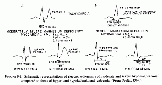
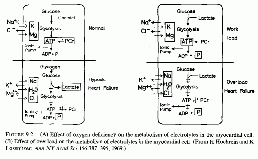
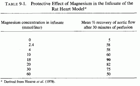
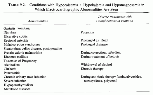
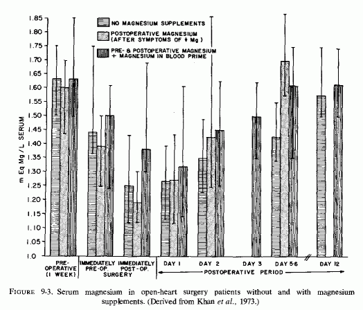
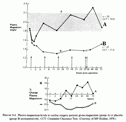
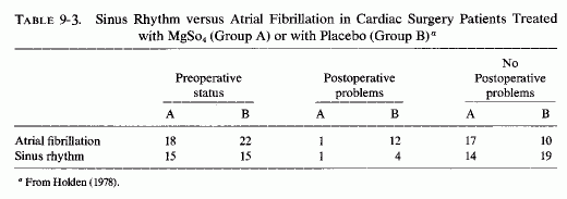
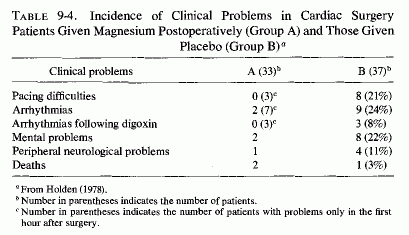
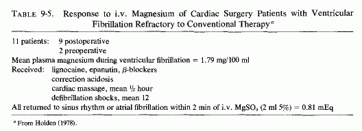
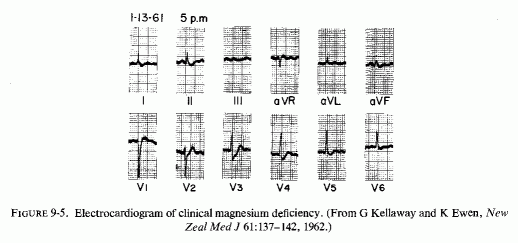
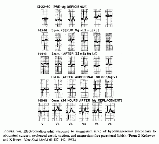
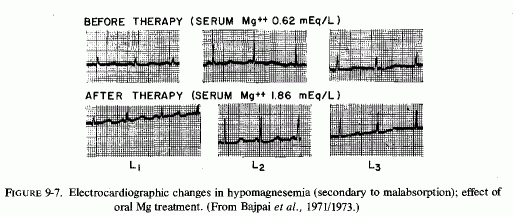
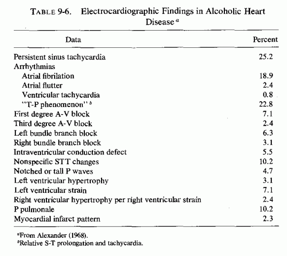
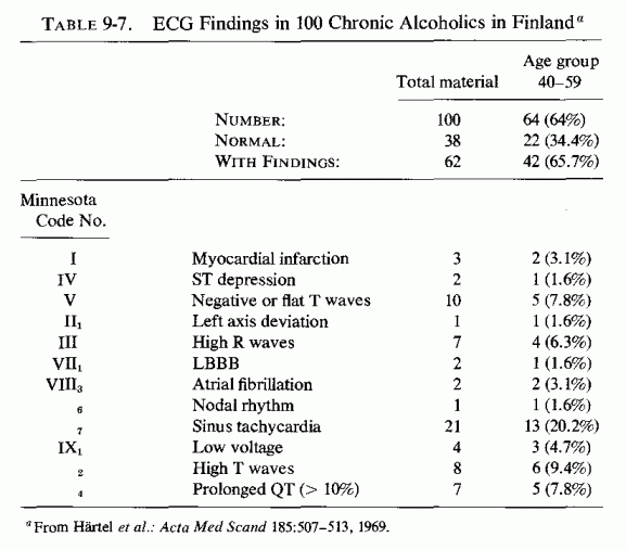
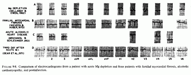
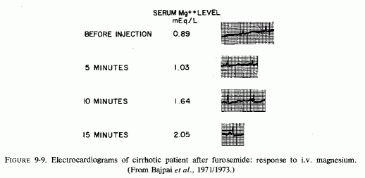
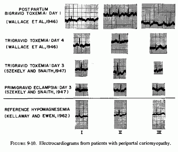